|

Synthesis and Spectroscopic Study of Benzanthrone 3-N-Derivatives as New Highly Fluorescent Dyes
E.M.Kirilova1, A.I.Puchkin1, I.D.Ivanova2, A.Ruža1, G.K.Kirilov, N.Orlova2
1Chemistry Department, Daugavpils University, 13 Vienibas str., LV-5401, Daugavpils (Latvia);
2Faculty of Chemistry, University of Latvia, 48 Valdemara str., LV-1013, Riga (Latvia).
e-mail: elena.kirilova@inbox.lv
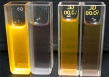
Today many techniques use fluorescent dyes for the labelling of biological objects. New practical uses call for the synthesis of new fluorescent probes with improved properties. In present work, we have synthesized a number of 3-aminobenzanthrone N‑derivatives – substituted amines, imines and amidines, obtaining highly fluorescent compounds [1]. Synthesized dyes are the analogues of cell membrane hydrophobic probe – 3‑methoxybenzanthrone, but new derivatives are long-wavelength light-emitting fluorescent dyes.
The spectral behaviour of the obtained dyes was investigated. We have studied the spectral properties of prepared derivatives – absorption and fluorescence spectra in various solvents. For obtained dyes large Stokes shift values (about 100 nm) are observed, excitation maxima of the dyes are located near 500 nm and emission maxima near 650 nm. In addition these compounds are showed strong fluorescent solvatochromism.
It was found that many of synthesized compounds are quite sensitive to the surrounding environments and are potential fluorescent probes for screening structural and functional alterations of cell membranes.

Synthesis and Crystal Structure of Novel Amidino Derivatives of Benzanthrone
E.M.Kirilova1, I.D.Ivanova2, A.I.Puchkin1, S.V.Belyakov3
1Chemistry Department, Daugavpils University, 13 Vienibas str., LV-5401, Daugavpils (Latvia);
2Faculty of Chemistry, University of Latvia, 48 Valdemara str., LV-1013, Riga (Latvia);
3Latvian Institute of Organic Synthesis, Riga, Latvia.
e-mail: elena.kirilova@inbox.lv
Today many techniques use fluorescent dyes for the labeling of biological objects [1]. Practical applications uses call for the synthesis of new fluorescent compounds with improved properties for a specific requirement. In this connection the intensive investigations for preparation of new fluorescent probes now have developed.
In our previous investigations were synthesized a number of benzanthrone N‑containing derivatives, obtaining highly fluorescent compounds [2, 3]. Synthesized dyes are sensitive long-wavelength light-emitting fluorescent dyes, which have high photostability and low cytotoxicity. It has been shown that some of obtained compounds are potential fluorescent probes for screening structural and functional alterations of cell membranes and for estimation of the immune state [2].
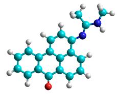
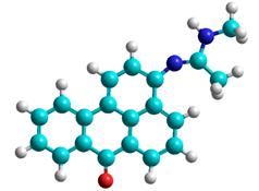
The aim of this work was the modification of some of benzanthrone derivatives (I, III) by including new substituents in chromophoric system in order to find potential fluorescent probes with large luminescence intensity and high stability. Here we present the synthesis of several new benzanthrone derivatives (II, IV) with amidine group:

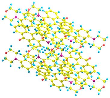
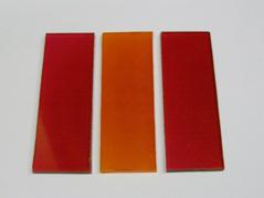
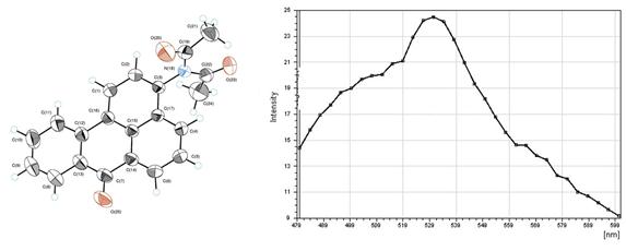
Synthesized derivatives have bright from yellow to red fluorescence in organic solvents and solid state. The structure of obtained compounds was confirmed by NMR and IR spectroscopy. In addition thermal analysis and crystal structures of studied compounds have been investigated.
1.Kirilova, E. M; Kalnina, I; Kirilov, G. K; Meirovics, I. J. Fluoresc 2008, 18 (3-4), 645-648.
2.Kirilova, E. M; Belyakov, S. V; Kalnina, I. Topics in Chemistry & Materials Science, Sofia: Heron Press (ed: G. Vayssilov, R. Nikolova) 2009, 3, 19-28.
3.Gorbenko, G.; Trusova, V.; Kirilova, E.; Kirilov, G.; Kalnina, I.; Vasilev, A.; Kaloyanova, S.; Deligeorgiev, T. Chem. Phys. Lett, 2010, 495, 275–279.
Synthesis of New Solvatochromic Dyes by Reduction of Benzanthrone Azomethines
I.D.Ivanova1, N.V.Orlova1, E.M.Kirilova2
1Faculty of Chemistry, University of Latvia, 48 Valdemara str., LV-1013, Riga (Latvia);
2Chemistry Department, Daugavpils University, 13 Vienibas str., LV-5401, Daugavpils (Latvia).
Many derivatives of benzo[de]anthracene-7-one exhibit strong emission, which accounts for their use in practice as active lasing media and fluorescent probes for investigation of biological objects. In present work a number of new derivatives of 3-aminobenzanthrone were synthesized. In summary, ten new dyes were synthesized in good yields (80-87%) via the reduction of corresponding azomethine derivative by sodium borohydride in DMF solutions.
The structure of obtained compounds was confirmed by NMR and FT-IR spectroscopy and mass spectrometry. Single-crystal structures of obtained dyes were determined by X-ray diffraction studies. In addition, thermal stability of the synthesized chromophores has been undertaken using TG–DTA. The influence of solvents with various polarities upon absorption and emission spectra was investigated. The absorption and luminescent spectra of the novel compounds in several solvents of different polarity were investigated. The synthesized dyes absorb at 520-580 nm with high extinction coefficients, have relatively large Stokes’ shifts (about 100 nm), and emit at 650-720 nm showing both absorption and fluorescence solvatochromism similarly to studied earlier 3-amino derivatives of benzanthrone [1,2]. The results indicated that these dyes were strongly dependent on solvents and show generally bathochromic shifts as the polarity of solvents was increased. These characteristics of obtained dyes demonstrate their potential as biomedical probes for proteins, lipids and cells.
1.Kirilova, E. M; Kalnina, I; Kirilov, G. K; Meirovics, I. J. Fluoresc 2008, 18 (3-4), 645-648.
2.Gorbenko, G.; Trusova, V.; Kirilova, E.; Kirilov, G.; Kalnina, I.; Vasilev, A.; Kaloyanova, S.; Deligeorgiev, T. Chem. Phys. Lett, 2010, 495, 275–279.
Novel Heterocyclic Imino and Amino Derivatives of Benzanthrone: Synthesis and Properties
I.D Ivanova1, N.V.Orlova1, A.I .Puchkin2, E.M.Kirilova2
1Faculty of Chemistry, University of Latvia, 48 Valdemara str., LV-1013, Riga (Latvia);
2Chemistry Department, Daugavpils University, 13 Vienibas str., LV-5401, Daugavpils (Latvia).
e-mail: elena.kirilova@inbox.lv
The design of new fluorescent molecules is of continuing interest for many applications in research and industry. Especially donor–acceptor p-conjugated organic materials have attracted considerable interests owing to their potential wide applications for development of photoactive materials. Many derivatives of benzo[de]anthracene-7-one are known as effective luminescent dyes with emission in the spectral region from green to red-purple, depending on the structure [1].
Recently we reported the synthesis, molecular structures and spectral properties of a series of amino, amidino, and iminobenzanthrones, which appeared to be particularly interesting because they lead to perspective luminescent materials [1]. The aim of this work is to create a series of benzanthrone amines and azomethines by including new heterocyclic substituents in benzanthrone chromophoric system in order to find novel luminophores with large luminescence intensity and high stability.
Target benzanthrone derivatives are synthesized by condensation reaction of corresponding aminobenzanthrone with appropriate heterocyclic aldehydes with following reduction of obtained imines by NaBH4 in DMF solution:
Synthesized derivatives have bright from yellow to red fluorescence in organic solvents and solid state. The structure of obtained compounds was confirmed by NMR, IR and mass spectroscopy. In addition thermal properties and crystal structures of prepared compounds have been investigated.
1.B.M. Krasovitskii, B.M. Bolotin, Organic luminescent materials. - VCH Publishers, 1988
2.E. M Kirilova, I. Kalnina, G. K. Kirilov, S. V Belyakov, I. Meirovics. Spectroscopic Study of Benzanthrone 3-N-Derivatives as New Hydrophobic Fluorescent Probes for Biomolecules// J.Fluoresc. – 2008 - Vol. 18 (3-4) – P.645-648
3. V.M Trusova, E. Kirilova, I. Kalnina, G. Kirilov, O.A. Zhytniakivska, P.V. Fedorov, G. Gorbenko, Novel Benzanthrone Aminoderivatives for Membrane Studies.// J.Fluoresc.- 2012 -Vol. 22 (3) – P.953-959
Structural Investigation of Novel Organic Materials with Photophysical Properties
S.Belyakov*1, A.Yanychev2, M.Fleisher1
1Latvian Institute of Organic Synthesis, Riga, Latvia;
2Riga Technical University, Riga, Latvia.
e-mail: serg@osi.lv
In recent years a series of organic compounds with photophysical properties has been studied by means of X-ray diffraction and theoretical calculations. The molecular and crystal structure of these compounds are intensively investigated, because there are much unexpected results. In this presentation the results of the following investigations will be reported: molecular and crystal structures of 2-(4’-Dimethylamino)-benzylydene-1,3-indandione (DMABI) analogues, structure of 8-mercaptoquinoline and 8-selenoquinoline complexes containing metal atoms, structure of benzanthrone derivatives. Part of these investigations has been published earlier as original articles [1-6]; the other part will be presented for the first time.
1. B Silina, E., Belyakov, S., Ashaks, J., Pecha, L., Zaruma, D. Tris(2-methylquinoline-8-selenolato-k1N,Se)antimony (III). Acta Crystallogr. 2007, vol.63.,Sec.C. p. m62-m64.
2. Belyakov, S., Kampars, V., Pastors, P.J., Tokmakov, A. 2-(4,5,6,7,8,9-Hexahydro-6a-azaphenylen-2-ylmethylene)indian-1,3-dione. Acta Crystallogr. 2008, vol.64.,Sec.E. p. o1200.
3. Kirilova, E.M., Belyakov, S.V., Kirilov, G.K., Kalnina, I., Gerbreder, V. Luminescent properties and crystal structure of novel benzanthrone dyes. J. Luminescence 2009, vol.129, p. 1827-1830.
4. Silina, E., Belyakov, S., Ashaks, J., Pecha, L., Zaruma, D. Crystal structure of catena-poly[bis (8-mercaptoquinolinato-N,S)disilver(I)], Ag2(C9H6NS)2. Zeitschrift für Kristallographie 2010, no.225. p. 211-212.
Thermal Properties of Chromium Complexes with N-benzanthronylamidines
A.I.Puchkin1, I.D.Ivanova2, E.M.Kirilova1
1Chemistry Department, Daugavpils University, 13 Vienibas str., LV-5401, Daugavpils (Latvia);
2Faculty of Chemistry, University of Latvia, 48 Valdemara str., LV-1013, Riga (Latvia).
e-mail: aleksandrs.puckins@du.lv
The chemistry of functional materials of metal complexes and their applications have been of great interest in recent years. Compounds containing amidine group have played important roles as ligands for various complexes of metals in organometallic chemistry. These systems have interesting chemical and spectral properties for practical applications. For example, the use of amidinate-ligated iridium complexes for fabrication of high efficiency phosphorescent organic light-emitting devices has been recently demonstrated [1]. Therefore it was of interest to prepare new complexes of transition metal ion and ligand containing electron-donating amidine group.
The object of the present research was to investigate the thermal behaviour of chromium (III) complexes incorporating earlier synthesized [2] N-benzanthronylamidine ligands and to characterize it by means of elemental analysis, infrared spectroscopy, and differential thermal analysis (TG-DTA). It was investigated coordination of fifteen amidines with Cr(NO3)3, obtaining the formation of seven complex. Elemental analysis data with the hexacoordination tendency of Cr3+ complexes, let us to suggest an octahedral geometry for obtained complexes with three molecules coordinated to the metal centre. The TG-DTA curves provided previously unreported information about the thermal stability and thermal decomposition of these compounds.
1. Y. Liu, K. Ye, Y. Fan et al. Chem. Commun. 25 (2009) 3699-3701.
2. E.M Kirilova, S. V Belyakov, I. Kalnina. in: G. Vayssilov, R. Nikolova (Eds), Topics in Chemistry & Materials Science, Sofia: Heron Press, 3, 2009 pp.19-28.
Spektrofluorometrisko pētījumu metožu pielietojums smago metālu jonu noteikšanai ūdens vidē
A.Pučkins1, E.Kirilova1
1Daugavpils Universitāte, Daugavpils, Latvija
e-mail: aleksandrs.puckins@du.lv
Vides piesārņošana ar smagajiem metāliem mūsdienās ir ļoti aktuāla problēma. Tāpēc pašlaik ir pievērsta liela uzmanība, lai izstrādātu jaunas jutīgas šo vielu noteikšanas metodes.Ņemot vērā zināmo faktu, ka metālu joni var ietekmēt fluorescentā savienojuma, ar kuru saistās, fluorescences parametrus, tika veikts pētījums, kura laikā tika noskaidrota spektrofluorometrisko metožu pielietojuma iespējamība smago metālu detektēšanai ūdens vidē.
|
|
|
Pētījumu laikā tika uzņemti vairāki krāsvielu fluorescences intensitātes spektri, pēc kuriem spriežot tika secināts, ka dažādu smago metālu klātbūtne var izraisīt dažādus efektus: gan organisko fluoroforu fluorescences pavājināšanu, gan pastiprināšanu.
Pētījuma gaitā tika noskaidrots, ka ar spektroflorometriskas metodes palīdzību var veikt gan kvalitatīvo, gan kvantitatīvo dažu smago metālu noteikšanu ūdens vidē.
Spektrofluorometrisko pētījumu metožu grupai ir raksturīga vesela virkne priekšrocību, kas nepiemīt pārējām analīzes metodēm un lietderīgi atšķir to no citām metodēm: zemas izmantošanas izmaksas, eksperimentu vienkāršums, metodes straujums un jūtīgums.
Amidines of Benzanthrone as Fluorescent Sensor for Metal Ions
A.I.Puchkin1, I.D.Ivanova2, E.M.Kirilova1
1Chemistry Department, Daugavpils University, 13 Vienibas str., LV-5401, Daugavpils (Latvia);
2Faculty of Chemistry, University of Latvia, 48 Valdemara str., LV-1013, Riga (Latvia).
e-mail: aleksandrs.puckins@du.lv
Pollution of the environment by toxic metals is a major problem in the last decays due to their increasing utilization in industry and agriculture.. These pollutants are discharged or transported into the atmosphere and aquatic and terrestrial environments and have the potential to reach high concentrations. Large concentrations of some metal ions have been shown to cause brain damage, and are potentially lethal. Due to potential health risks associated with exposure to metals, legal constraints on the allowed abundance of metals are becoming increasingly strict.
As a result of the health threats posed by some metals, new methodologies that allow for their simple detection and quantitation are of interest. Today many fluorescent organic sensors were designed for recognition and detection of heavy and transition metal ions. These chemosensors are compounds that incorporate a binding site, a fluorophore, and a means of communication between the two. The interaction of a metal ion with an organic ligand may result in fluorescent enhancement or fluorescence quenching.
Our recent efforts have been focused largely on synthesizing novel organic fluorophores such as N-containing benzanthrone derivatives [1,2]. The present work focuses on the spectral characterization of benzanthronylamidines and its complexes with transition metal ions. Obtained results testify that investigated dyes can be used as sensitive fluorescent chemosensors for transition metals: the one of them displays positive response (3-7-fold fluorescence increasing) to the presence of Ni2+ or Co2+ ions, but some synthesized amidines show negative response (quenching) in fluorescence intensity to Cr ions. Further investigations can lead to development of new method for the detection of some cations.
1. E.M Kirilova, S. V Belyakov, I. Kalnina. in: G. Vayssilov, R. Nikolova (Eds), Topics in Chemistry & Materials Science, Sofia: Heron Press, 3, (2009) 19-28.
2. V.M. Trusova, E. Kirilova, I. Kalnina, G. Kirilov, O.A. Zhytniakivska, P.V.Fedorov, G. Gorbenko. J.Fluoresc. 22 (2012) 953-959.
Jaunās, uz 3-aminobenzantrona bāzes, sintezētās krāsvielas
A.Ruža1, A. Volkova1, E.M.Kirilova1,
1Ķīmijas fakultāte, Daugavpils Universitāte, Vienības iela 13., LV-5401, Daugavpils (Latvija)
e-mail: elena.kirilova@inbox.lv; anita.ruza@gmail.com
Benzantrons un tā atvasinājumi tiek plaši izmantoti dažādās zinātnes sfērās. Tā krāsvielas ir labi zināmi luminofori, kam piemīt izteikta krāsa un fluorescences intensitāte, tās ir stabilas un gaismas ietekmē nedegradējas. Ar benzantrona krāsvielām patreiz var iekrāsot gan ikdienā izmantojamas lietas (piem., audumus), gan dažādus polimērus un pat izmatot tās LCD dienas gaismas spuldzītes un lāzeros.
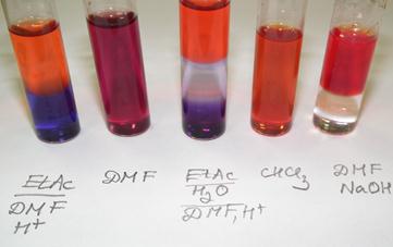
Krāsvielu sintēzei nepieciešamā 3-NH2-BA iegūšana notiek divos posmos: benzantrona nitrēšana ar turpmāku reducēšanu Na2S klātbūtnē. Amidīnu sintēzes process ir sekojošs: 3-NH2-BA kopā ar attiecīgo amīdu vāra kopā ar POCl3 katalizatoru ~ 90 – 100oC. Vēlāk reakcijas maisījumu neitralizē ar piesātinātu amonjaka šķīdumu un izkritušās nogulsnes nofiltrē. Parasti pēc to izžūšanas ir nepieciešama nogulšņu pārkristalizācija, lai atbrīvotos no neorganikas piemaisījumiem un varētu izaudzēt vielu kristālus. Papildus tiek iegūti arī amidīni ar broma atomu, kas piesaistīts pie 2. vai 3. benzantrona oglekļa atoma. Broma klātbūtnē mainās krāsvielu vizuālais izskats, luminescences krāsa un spektrālās īpašības.
Spektrāliem pētījumiem tiek sagatavoti amidīnu šķīdumi dažādos organiskajos šķīdinātājos (DMF, CHCl3, u.c.) koncentrācijā 10-4 M. Daļai no iegūtajām krāsvielām tiek pārbaudīta to spēja veidot kompleksos savienojumus ar smagajiem metāliem.
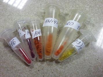
Pašlaik jau ir sintezētas 36 krāsvielas un ir plānots iegūt vēl vismaz sešas. No esošajām krāsvielām, visizteiktākā luminescence sausā veidā ir savienojumiem AM16, AM20 un AM22.
Thin Films Fabricated from Benzanthrone Luminescent Dyes
G.K.Kirilov1, A.S.Bulanov1, M.Fleisher2, E.M.Kirilova1, I.Mihailova1, S.Gonta2
1Daugavpils University, Vienibas 13, LV-5401, Daugavpils, Latvia
2Institute of Microbiology and Biotechnology, University of Latvia, Kronvalda blvd. 4, Riga LV-1586, Latvia
e-mail: georgk@inbox.lv
Organic thin films are used in a number of scientific and technical applications one of which is producing cheap and large scale electronic and optical devices [1,2]. Thin films samples of novel organic luminophores - 3‑N‑(N’-methylacetamidino)benzanthrone and 3‑N‑(N`,N`‑dimethylbenzamidino)benzanthrone deposited on glass substrate were prepared by thermal evaporation obtaining thin films of 2.5 to 3 µm thickness.
The structural and spectroscopic properties of obtained films were investigated by confocal microscope with input of femtosecond laser radiation, X-ray diffractometer and scanning electron microscope. In addition, quantum chemical calculations were performed, indicating electron properties of studied dye molecule in ground and excited state. Heats of formation, dipole moments and other parameters were extracted directly from the data files following the geometry optimizations. The calculations show a planar configuration for the aromatic core of these compounds and two possible orientations of amidine substituents.
|
E= -4468.70 (kcal/mol) |
E= -4455.22 (kcal/mol) |
1.J. R. Sheats, H. Antoniadis, M. Hueschen, W. Leonard, J. Miller, R. Moon, D. Roitman, A. Stocking, Organic Electroluminescent Devices, Science, 273 (1996) 884-888.
2.B.M. Krasovitskii, B.M. Bolotin, Organic luminescent materials. VCH Publishers, 1988.
Luminescence and Structural Properties of Benzanthrone Dyes Thin Films
I.Mihailova1, G.Kirilov1, A.Bulanovs1, E.Kirilova2
1Innovative Microscopy Center, Daugavpils University, Daugavpils, Latvia.
2Department of Chemistry, Daugavpils University, Daugavpils, Latvia.
e-mail: irena.mihailova@du.lv
We report luminescence and structural properties of 3-N, N-diacetylaminobenzanthrone thin films deposited on glass substrate by thermal evaporation. The optical and structural properties of organic thin films were studied by means of the confocal microscope with an input of femtosecond laser radiation, X-ray diffractometer, and scanning electron microscope (SEM).
Intense luminescence with the maximum at 530 nm was observed when excited by laser radiation with the wavelengths 458-514 nm. In addition, the luminescence caused by two-photon absorption of infrared femtosecond (fs) laser radiation has been investigated. The study of the structure of benzanthrone derivative thin films, created by X-ray diffraction (XRD) methods, indicate the distance between molecular layers and ordered molecular fragments.
It was found that prepared films of 3‑N,N‑diacetylaminobenzanthrone are highly ordered materials with molecular layers; X-ray diffraction analysis indicate that the distance between these layers is ~6.5 Å. Similar ordering this compound shows in monocrystal packing.
Application of Fluorescent Pigments and Dyes in the Research of Plant Stem Cells and Programmed Cell Death
M.Selga*1, T.Selga2, A.Ozolina1
1Daugavpils University, Daugavpils, Latvia;
2Faculty of Biology, University of Latvia, Riga, Latvia
E-mail: turs.selga@lu.lv
Different types of stem cells have been stated both in plants and animals [1]. The research of stem cells particular abilities, regulation and fate becomes increasingly important due to the fast development of regenerative medicine and new methods of plant propagation. However little is known about the flowering plant stem cell diversity, pools, developmental strategies and stemness of differentiated cells and regulation of programmed cell death [2]. Cells of tobacco (Nicotiana tabacum L.), field beans (Vicia faba L.) and other plants cultivated in optimal conditions were researched by the light and confocal laser-scanning microscopy. We have stated that:
· The asymmetric strategy of stem cell division and fate of daughter cells;
· Switching the structural mechanisms of mitosis that correlates with the reflective processes of cell differentiation and dedifferentiation;
· The lasting association of subordinated unequal daughter nuclei and cells via plasmodesmata due to arrested asymmetric mitosis;
· Both mitosis and asymmetric programmed cell death is double regulated – by intrinsic and extrinsic signalling via evident structural pathways;
· Plastid-nucleus complex encircles mitosis in photosynthesising cells and exists during programmed cell death [3].
Peculiarities of staining with fluorescent dyes in different stages of cell development will be discussed.
1.Lin, H. Cell Biology of Stem Cells: an Enigma of Asymmetry and Self-renewal. J Cell Biol, 2008, vol.180, p 257-260.
2.Krishnamurthy K.V., Krishnaraj R., Chozhavendan R. et al The programme of cell death in plants and animals – A comparison. Current Sci. 2000. vol. 79, p. 1169-1181.
3.Selga T., Selga M., Gobins V., et al. Plastid-nuclear complexes: permanent structures in photosynthesizing tissues of vascular plants. Env and Exp Biol, 2010, vol. 8, p. 85–92.
The Influence Of High Temperature On Oxidative Processes and Accelerated Senescence in Wheat Seedling (Triticum aestivum L.) Coleoptile
M.Savicka*1, N.Škute1
1Institute of Ecology, Daugavpils University, Daugavpils, Latvia
e-mail: marina.savicka@du.lv
Most environmental stresses are affecting on the production of active oxygen species in plants, causing oxidative stress. Temperature, along with light, humidity, etc., belongs to the important factors in the environment of plants. It is also a tool for research, for in many cases the processes of growth can be differentiated by their temperature responses. Heat shock is a form of the oxidative stress, resulting in the formation of many toxic ROS in plants (Tsang et al. 1991; Bhattacharjee, Mukherjee, 2003). High temperature induced oxidative stress led to accelerated senescence of plants. Therefore, the objective of this work was to study the response of coleoptile of wheat seedlings to elevated temperatures. The present study investigated the effect of heat stress on different stages of growth in wheat (Triticum aestivum L.). The behaviour of wheat seedling coleoptile was studied, particularly at the physiological level. High temperature is known to induce oxidative injury in plants by inducing production of active oxygen species. Active oxygen species, cause lipid peroxidation (LP) and consequently membrane injury. High-temperature (15, 30 and 60 min and 24h at 42°C) influence on senescent organ of wheat seedlings (Triticum aestivum L.) was analyzed taking into consideration the changes in some changes on the molecular level, such as content of LP products (malodialdehyde (MDA) and conjugated dienes (CD)) and membrane permeability. The effect of high temperature was analyzed at the early stages (4 day-old) and at the late stages (7 day-old) of seedling development. LP level was directly correlated with temperature, exposure time, and their interaction. Long-term high-temperature exposure (24h at 42°C) significantly increased LP (MDA and CD content) in coleoptile of wheat seedlings at 4 and 7d of seedling development. However, the increase in LP products was observed only at the late stages of seedling development after short-term high temperature exposure. Heat shock caused an increase in the electric conductivity of cell membrane, which led to the loss of membrane selective permeability.
1.Tsang, E.W.T., Bowler, C., Hérouart, D., Van Camp, W., Villarroel, R., Genetello, C. Differential regulation of superoxide dismutases in plants exposed to environmental stress. Plant Cell. 1991, 3: 783–792.
2.Bhattacharjee, S., Mukherjee, A.K. Implications of reactive oxygen species in heat shock induced germination and early growth impairment in Amaranthus lividus L. Biologia Plantarum. 2003, 47 (4): 517-522.
Application of New Fluorescent Dyes ABM, AM1, AM15 and P8 in Plant Cell Biology. Problems and Perspectives.
A.Ozolina1, T.Selga2, M.Selga1
1Daugavpils University, Daugavpils, Latvia;
2Faculty of Biology, University of Latvia, Riga, Latvia
e-mail: turs.selga@lu.lv
To find out possible application of new stains ABM, AM1, Am15 and P8 in plant cell biology we used different fixation and staining techniques in different combinations. Reliability of results were analysed with bright field and confocal laser-scanning microscopy.
For bright field microscopy bulbs of onion Allium cepa L. were dissected and epidermis from different layers of leaf scales was removed. Epidermal peels from the middle of tobacco, pelargonium leaves and the outer, middle and inner layer of leaf scales of onion bulbs were stripped and transferred to droplets of aceto-ethanol on microscopic slides, washed and contrasted with aceto-orcein or methylene blue. Mesophyll was fixed with 2% glutaraldehyde in sodium cacodylate buffer or in aceto-ethanol, washed with water, frozen in a droplet of water, cut by razor under 50x magnification; and cuttings were contrasted on microscopic slides with aceto-orcein. Leaf pieces of mesophyll were excised in the middle of the leaf blade among large veins, soaked in aceto-ethanol for, rinsed in dist. water, maturated in 1M HCl at 600 C, rinsed in dist. water, contrasted with aceto-orcein or methylene blue, placed on microscopic slides, covered with cover slips, and gently crushed by pressure. Largely or partly separated palisade parenchyma cells due to destruction of cellulose were examined.
Samples were examined under bright field illumination under a light microscope LEICA DM 2000. Photographs were taken with a digital camera Leica EC3 and images processed with LAS EZ and Paint Shop Pro 4.
For confocal laser-scanning microscopy to observe plant tissue in vivo samples were mounted in tap water on a glass depression slide and placed under a cover slip. To limit water evaporation and cover slip drift during long-term observations, cover slip was attached to glass slide with an adhesive tape.
Smples were fixed with 2% glutaraldehyde in sodium cacodylate buffer or in aceto-ethanol and washed with water. Samples were stained with propidium iodine, bodipy, mitotracker green, ABM, P8, AM1 or AM15 in different combinations.
Laser scanning confocal microscopy was conducted with a Leica DM RA-2 microscope equipped with a TCS-SL confocal scanning head (Leica Microsystems, Bannockburn, Ill.). Images were collected with a Leica 40x HCX PL FLUOTAR objective (NA=0.75) and 100× HCX PlAPO oil immersion objective (NA=1.40). Pictures were recorded by taking z-stacks that captured multiple organelles. At least three such z-stacks were taken and analyzed for each sample. We used 0.3 μm thick optical sections and collected from 1 to 10 μm thick samples.
AM1,AM 15, P8 and ABM were excited with a 488-nm band of a four-line argon ion laser. Fluorescence was detected between 530 nm and 580 nm. GFP was excited with a 488-nm band of a four-line argon ion laser. Chlorophyll fluorescence was excited at 633 nm. GFP fluorescence was detected between 500 nm and 560 nm. Chlorophyll fluorescence was detected between 680 nm and 700 nm.
Mycobacterium tuberculosis antibiotiku rezistenci izraisošo mutāciju noteikšana pēc DNS denaturācijas laikā novērotajām fluorescences līmeņa izmaiņām
A.Kalviša1, I. Jansone1, V. Baumanis1
1Latvijas Biomedicīnas pētījumu un studiju centrs, Rīga, Latvija
e-mail: adrija.kalvisa@gmail.com
Ievads: Ar rezistenci saistīto punktveida mutāciju spektrs un izvietojums M. tuberculosis genomā ir plašs, tādēļ ir nepieciešama metode ātrai mutāciju klātbūtnes konstatēšanai vairākos genoma fragmentos vienlaicīgi. Augstas izšķirtspējas DNS kušanas līknes analīze (high resolution melting jeb HRM) ir metode, kas pēc fluorescences nosaka mutāciju izraisītas DNS fragmentu denaturācijas īpašību izmaiņas. HRM izmantošana iepriekš veiksmīgi izmantota, nosakot mutācijas pēc DNS, kas izdalīta no M. tuberculosis tīrkultūrām1, tomēr nav skaidras metodes pielietošanas iespējas atšķirīgas kvalitātes klīnisko paraugu analīzē.
Mērķis: Noskaidrot HRM metodes pielietošanas iespēju robežas M. tuberculosis antibiotiku rezistences mutāciju molekulārģenētiskajai noteikšanai no klīniskā materiāla izdalītos DNS paraugos. Pārbaudīt metodes jutību genoma fragmentos ar atšķirīgiem mutāciju veidiem un mainīgu mutāciju daudzveidību.
Metodes: Darbā tika izmantota M. tuberculosis DNS, izdalīta no slimnieku klīniskā materiāla. Pētījuma objekti:
a) ar streptomicīna rezistenci saistītās mutācijas 16S rRNS kodējošā rrs gēna fragmentā 18 paraugos (2 dažādas mutācijas 8 paraugos, 10 savvaļas tipa paraugi).
b) GyrA gēna hinolonu rezistenci determinējošā rajona (QRDR) mutācijas 32 paraugos (8 dažādas mutācijas 13 paraugos, 8 savvaļas tipa paraugi un 3 jaukta tipa paraugi).
DNS fragmenti pavairoti ar reālā laika PCR (polimerāzes ķēdes reakcijas) iekārtu Viia7 (LifeSciences) DNS interkalējošas fluorescentās krāsas klātbūtnē. Katrs paraugs amplificēts divās paralēlās reakcijās. Mutāciju klātbūtne konstatēta, mērot interkalējošas krāsas fluorescenci pakāpeniskā temperatūras pieaugumā un salīdzinot amplikona denaturācijas temperatūru (TD) ar atskaites DNS fragmenta TD. Visi iegūtie dati par mutāciju klātbūtni salīdzināti ar sekvenēšanas rezultātiem.
Rezultāti: a) HRM analīze rrs gēna 8/8 mutantiem paraugiem un 7/10 savvaļas tipa paraugiem sakrita ar sekvenēšanas rezultātiem, savukārt atlikušie 3 savvaļas tipa paraugu dati bija pretrunīgi.
b) GyrA gēnam sakrita tikai 3/13 savvaļas tipa un 1/8 mutanto paraugu piederība, bet 3 jaukta tipa paraugi deva pretrunīgus rezultātus.
Secinājumi: HRM metode veiksmīgi izmantota streptomicīna rezistences mutāciju konstatēšanai un atšķiršanai no neitrālām mutācijām. Savukārt liela mutāciju daudzveidība un mutācijas, kuru ietekme uz TD ir maza, apgrūtina mutāciju atšķiršanu un HRM izmantošanu hinolonu rezistences noteikšanai klīniskajos paraugos.
1. Chen, X., Kong, F., Wang, Q., Li, C., Zhang, J., Gilbert, G.L. Rapid detection of isoniazid, rifampin, and ofloxacin resistance in Mycobacterium tuberculosis clinical isolates using high resolution melting analysis. J Clin Microbiol, 2011, vol. 49, p. 3450-7
Melanokortīna 1. receptora gēna polimorfismu funkcionālās lomas izvērtēšana, izmantojot konfokālo lāzerskenējošo mikroskopiju
A.Ozola*1, R.Petrovska1, I.Mandrika1, O.Heisele1, D.Pjanova1
1Latvijas Biomedicīnas pētījumu un studiju centrs, Rīga, Latvija
e-mail: aija.ozola@biomed.lu.lv
Ievads. Melanoma ir ļaundabīgs ādas audzējs, kas veidojas no melanocītiem. Viens no zināmākajiem melanomas riska gēniem ir melanokortīna 1. receptora gēns (MC1R), kas iesaistīts ķermeņa pigmentācijas regulācijā. MC1R ir ļoti polimorfs. Asociāciju pētījumos ir parādīta biežāk sastopamo MC1R polimorfismu saistība ar ķermeņa pigmentāciju un/vai paaugstinātu melanomas risku, savukārt in vitro pētījumos ir pierādīta to ietekme uz receptora funkcionālo aktivitāti. Pārējie MC1R polimorfismi ir reti sastopami, un tāpēc tiek uzskatīts, ka to ietekme uz melanomas risku nav vērā ņemama. Tomēr daļa no šiem polimorfismiem varētu mainīt receptora funkcionālo aktivitāti un līdz ar to arī ietekmēt melanoma risku.
Mērķis. Izvērtēt Latvijas populācijā atrasto reto MC1R polimorfismu (g.133T>C Phe45Leu, g.248C>T Ser83Leu, g.265G>C Gly89Arg, g.284C>T Thr95Met, g.363C>G Asp121Glu, g.493G>A Val165Ile, g.550G>C Asp184His, g.562G>A Val188Ile, g.637C>T Arg213Trp) ietekmi uz receptora funkcionālo aktivitāti.
Metodes. Izmantojot PCR mutaģenēzi, tika izveidoti MC1R konstrukti ar minētajiem polimorfismiem. Mikroskopijas vajadzībām tika izveidots otrs MC1R konstruktu komplekts, kurā polimorfismus saturošie MC1R varianti tika sajūgti ar GFP (green fluorescent protein). Visi konstrukti tika ekspresēti BHK (baby hamster kidney) šūnās. Konstrukti bez GFP tika izmantoti cikliskā adenozīna monofosfāta (cAMP) aktivācijas līmeņa noteikšanai. Konstrukti ar GFP tika analizēti, izmantojot konfokālo lāzerskenējošo mikroskopu Leica TCS SP2. Šūnām, kas tika analizētas mikroskopijā, membrānas tika iekrāsotas ar fluorescento krāsvielu Alexa FluorÒ 633 WGA (wheat germ agglutinin). MC1R konstruktu šūnas virsmas ekspresijas līmeņa kvantitatīvai analīzei tika izmantots programma Leica Confocal Software (Las AF v2.6.0). Konfokālās mikroskopijas attēlos katrai no kopumā sešām šūnām (pa trim šūnām no divām neatkarīgām transfekcijām) tika nejauši izvēlēti 10 lineāri ROI (regions of interest). ROI krustpunktā ar šūnas membrānu tika nolasīta GFP/WGA fluorescences intensitātes attiecība. Rezultātā katra konstrukta gadījumā tika iegūta datu kopa, kas sastāv no 120 GFP/WGA vērtībām. Iegūtās GFP/WGA vērtības tika izmantotas kvantitatīvā receptora šūnas virsmas ekspresijas līmeņa novērtēšanā, izmantojot neparametrisko Kruskal-Wallis testu un Dunn’s daudzkārtējo salīdzinājumu testu.
Rezultāti. Konfokālās lāzerskenējošās mikroskopijas attēlu analīze rāda, ka polimorfismu Ser83Leu un Gly89Arg gadījumā MC1R ekspresija uz šūnas virsmas salīdzinājumā ar savvaļas tipa receptoru tiek pilnībā kavēta, Asp121Glu un Arg213T gadījumā virsmas ekspresija ir pazemināta, bet pārējo polimorfismu gadījumā tā ir tādā pašā līmenī kā savvaļas tipa receptoram. Matriksa tabula, kas tika izveidota, izmantojot ranga summas no Dunn’s daudzkārtējo salīdzinājumu testa, rāda, ka ranga summu atšķirības starp polimorfajiem MC1R variantiem ar Ser83Leu, Gly89Arg, Asp121Glu vai Arg213T un savvaļas tipa receptoru ir statistiski ticamas (p<0.01). Cikliskā adenozīna monofosfāta līmeņa mērījumi uzrādīja, ka Gly89Arg pilnībā nomāc cAMP aktivāciju, Ser83Leu to ievērojami pazemina, bet pārējo polimorfismu gadījumā cAMP līmenis dažādās pakāpēs paaugstinās.
Secinājumi. Šis pētījums parāda, ka arī retie MC1R polimorfismi var ietekmēt receptora funkcionālo aktivitāti.
Pateicamies Dr. biol. Dāvidam Fridmanim par palīdzību statistiskajā analīzē.
Resonance Energy Transfer Study of Lipid Bilayer Interactions of Apolipoprotein A-I Variants
G.Gorbenko*1, V.Trusova1, E.Adachi2, C.Mizuguchi2, H.Saito2
1V.N. Karazin Kharkov National University, Kharkov, Ukraine;
2Institute of Health Biosciences, The University of Tokushima, Tokushima, Japan
e-mail: galyagor@yahoo.com
Due to its explicit distance dependence fluorescence resonance energy transfer (FRET) has long been used for obtaining structural information on a wide variety of biomolecular assemblies. In the present study this technique was employed to gain further insights into lipid interactions of apolipoprotein A-I (apoA-I) variants. ApoA-I is the main protein component of high-density lipoproteins promoting cholesterol efflux from plasma membrane and modulating phospholipid metabolism. Moreover, specific variants of human apoA-I, particularly those having G26R substitution mutation, are capable of forming amyloid fibrils associated with renal or liver failure. The N-terminal fragment (amino acids 1-83) has been identified as the predominant form of apoA-I in amyloid fibril deposits. This fragment contains three tryptophan residues, Trp8, Trp50 and Trp72 which have been recruited in this work as energy donors for membrane fluorescent probes 4-dimethylaminochalcone (DMC) and 3-methoxybenzanthrone (MBA), located in the interfacial bilayer region. Small unilamellar vesicles were prepared by extrusion technique from egg phosphatidylcholine (PC) and its mixture with cholesterol(Chol) (30 mol%). Shown in Fig. 1 are typical FRET profiles obtained for 5 apoA-I 1-83 fragments, referred to here as apoA83 (without mutation), G26R, G26R/W8F_W50F, G26R/W8F_W72F and G26R/W50F_W72F.
|
|
|
|
Fig. 1. FRET profiles for Trp-DMC donor-acceptor pair in PC (A) and PC/Chol (B) liposomes |
|
FRET data have been quantitatively interpreted in terms of the model of energy transfer in two-dimensional systems allowing for distance dependence of orientation factor. It was found that in PC membranes Trp residues adopt interfacial bilayer location, while in PC/Chol membranes they tend to reside in the aqueous phase. These findings support the hypothesis that Chol can affect the curvature of lipid-water interface in such a manner that thermodynamically favorable location of apoA-I becomes less buried.
This work was supported by the grants from European Social Fund (project number 2009/0205/1DP/1.1.1.2.0/09/APIA/VIAA/152) and Fundamental Research State Fund of Ukraine (project number F.41.4/014).
Styryl Derivatives as a New Probes For Yeast Cell Vitality Determination
I.P.Goriacha1, T.S.Dyubko1, V.D.Zinchenko1, I.V.Govor 2, L.D.Patsenke2, T.Deligeorgiev3, S.Kaloyanova3, N.Lesev3
1Institute for Problems of Cryobiology and Cryomedicine of the National Academy of Sciences of Ukraine, Kharkiv, Ukraine;
2State Scientific Institution "Institute for Single Crystals", Kharkiv, Ukraine;
3University of Sofia, Faculty of Chemistry, Sofia, Bulgaria.
e-mail: irynagor@gmail.com
Solutions for a number of biological, biophysical and cryobiological tasks is necessary to determine the viability of the cells in response to various factors cryobiological. In this work, we investigated the sensitivity of styryl fluorescent derivatives (Table) to a state of yeast cells Saccharomyces cerevisiae in the native state, after boiling, after the action of high doses of ozone (5 mg / l), and after the action of moderate doses of ozone (0,16, 0,48 and 0,80 mg / L) to determine the sensitivity to the listed factors. Used by flow cytometry and fluorescence spectroscopy.
table
|
Name |
Formula |
λmax excitation, nm |
λmax fluor., nm |
Quantum yield, [%] |
|
11-ST |
|
410 |
- |
81 |
|
16-ST |
|
510 |
- |
65 |
|
17-ST |
|
450 |
618 |
78 |
|
22-ST |
|
370 |
490 600 |
90 |
|
23-ST |
|
445 |
520 610 |
75 |
|
24-ST |
|
460 |
560 |
95 |
|
26-ST |
|
460 |
535 |
81 |
|
30-ST |
|
300 |
525 |
75 |

Found that the probes 17-ST, 24-ST, 26-ST and 30-ST are sensitive to the action of boiling and high doses of ozone. Probes 22-ST, 23-ST and 30-ST are sensitive to different moderate doses of ozone. Presumably, we attribute this to the sensitivity of dyes to study cell membrane potential, as well as to their ability to be integrated into the cell membrane and penetrate into the cells.
Monitoring of Free Radical Processes in Protein-lipid Systems with a New
Squaraine Dye
O.Kutsenko*1, V.Trusova1, G.Gorbenko1, T.Deligeorgiev2, E.Slobozhanina3, L.Lukyanenko3, G.Zubritskaya3
1V.N. Karazin Kharkov National University, Kharkov, Ukraine;
2Faculty of Pharmacy and Chemistry, University of Sofia, Sofia, Bulgaria;
3Institute of Biophysics and Cell Engineering, National Academy of Sciences of Belarus, Minsk, Belarus
e-mail: olzk@mail.ru
Amyloids are highly ordered protein aggregates whose deposition in organs and tissues is one of the characteristics of so called protein misfolding diseases. These disorders include Parkinson, Alzheimer, Huntington, prion diseases, etc. It was demonstrated that one of the reasons of amyloid cytotoxicity involves interaction of aggregated proteins with cell membranes, particularly with red blood cell membrane. The mechanisms of erythrocyte injury include modification of membrane fluidity, alterations in enzyme activity and oxidation state, apoptosis and oxidative stress.
The present study was undertaken to explore amyloid effect on membrane lipid oxidation. To this end, we used model systems containing lysozyme amyloid fibrils (Lz), hemoglobin (Hb) and liposomes from phosphatidylcholine (PC) and its mixtures with cardiolipin (CL) and cholesterol (Chol). For identification of the presence of active oxygen species novel squaraine dye SQ-1 was applied [1]. It has been previously found that Hb-induced formation of free radicals leads to the drastic quenching of SQ-1 fluorescence, resulting presumably from the reaction between reactive oxygen species and SQ-1 squaric moiety [2]. Lz association with lipid bilayers was not followed by SQ-1 spectral changes indicating insensibility of this probe to Lz-induced membrane modification. Hb addition to liposomes without Lz was followed by significant decrease of SQ-1 emission indicating the formation of free radicals. SQ-1 fluorescence quenching was found to depend on model membrane composition. In PC and PC/Chol bilayers the decrease of SQ-1 fluorescence was less pronounced than in CL-containing membranes. Interaction of the Hb with negatively charged membranes can be followed by protein unfolding, heme displacement and dissociation of heme-globin complex. This facilitates heme-lipid interaction and consequent iron-induced formation of free radicals leading to the increase of free radical formation in CL bilayers as compared to PC membranes. Cholesterol prevents Hb penetration into the inner membrane regions, thus inhibiting oxidative processes. Interestingly, lysozyme fibrils suppressed SQ-1 fluorescence quenching in all types of lipid bilayers, with the magnitude of this effect being most pronounced in CL-containing membranes. There are at least two explanations for Lz antioxidant effect. First, protein molecular groups can be the scavengers for free radicals. On the other hand, Hb and Lz can compete for the membrane binding sites, thereby reducing Hb penetration into the membrane interior and formation of free radicals.
This work was supported by the grant from Fundamental Research State Fund of Ukraine (project number F41.1/014) and Fundamental Research State Fund of Belarus Republic (project number B11K-152).
1.Ioffe, V.M., Gorbenko, G.P., Deligeorgiev, T., Gadjev, N., Vasilev, A. Fluorescence study of protein-lipid complexes with a new symmetric squarylium probe. Biophys. Chem., 2007, vol. 128, p. 75–86.
2.Trusova, V.M., Gorbenko, G.P., Deligeorgiev, T., Gadjev, N., Vasilev, A. A Novel Squarylium Dye for Monitoring Oxidative Processes in Lipid Membranes. J. Fluoresc., 2009, vol. 19, p. 1017–1023.
Studying Zinc Biology with Fluorescent Probes
E.I.Slobozhanina*1,Y.M.Harmaza1, N.M. Kozlova1, A.V.Tamashevski1
1Institute of biophysics and cell engineering of NAS B, Minsk, Belarus
e-mail: slobozhanina@ibp.org.by
Background: Zn2+ is essential trace element that is required for a wide range of biological processes, including cell proliferation and differentiation. The physiological concentration of Zn2+ in serum is 2–15 μM, when free Zn2+ level in most cells is extremely low (<1 nM). Although the cellular zinc levels are regulated by two families of specific transport proteins (ZnT and ZIP families) [1], intracellular Zn2+ homeostasis is sensitive to pathophysiological environmental changes [2].We have previously shown that exposure human erythrocytes to zinc sulphate in vitro led to accumulation of Zn2+ in the cells, increase in SH-, NH2-groups levels on the membrane surface and modification physical state of phospholipids and lateral distribution of membrane cholesterol [3, 4]. The aim of this work is clearing of interaction of Zn2+ homeostasis with induction of erythrocyte programmed cell death (eryptosis).
Methods: Human blood was obtained from normal donors. For detection of the intracellular labile Zn2+ level used FluoZin-3-AM/TS (Molecular Probes). The percentage of “eryptotic” cells was assessed by phosphatidylserine exposure using Annexin V-FITC (Beckman Coulter). Cytosolic esterase activity was assessed using Calcein-AM (Sigma). The formation of free radical compounds was evaluated using 2',7'-dichlorodihydrofluorescein diacetate (H2DCF-DA, Sigma). Investigations were performed on the spectrofluorimeter “Cary Eclipse” (Varian) and FACScan (BD).
Observations: Using specific ionophore, intra- and extracellular zinc chelators was determined the existence of the specific receptors on the erythrocytes surface and cellular stores responsible for the maintenance of zinc homeostasis. At the same time was shown that increase of intracellular labile Zn2+ level over 100 nM results in induction of eryptosis and Zn2+ cytotoxic effects (cytosolic esterase activity inhibition, phosphatidylserine redistribution on the cell membrane surface) is caused by intracellular molecular mechanisms lead to Zn2+ release from cellular stores. Also was established that the disturbance in "prooxidant/antioxidant" balance is possible trigger of eryptosis initiation.
Conclusion: The received results testify about existence of regulation mechanisms of zinc homeostasis in human erythrocytes and the fine concentration “border” between essential and toxic properties of Zn2+, failure of which can lead to the triggering of the processes of programmed cell death.
1.Eide, D.J. Zinc transporters and the cellular trafficking of zinc. Biochim. Biophys. Acta, 2006, vol. 1763, p. 711-722.
2.Tubek, S., Grzanka, P., Tubek, I. Role of zinc in hemostasis: a review. Biol. Trace Elem. Res, 2008, vol. 121, № 1, p. 1-8.
3.Harmaza, Y.M., Kozlova, N.M., Slobozhanina, E.I. Effects of zinc ions on the modification of human erythrocytes membranes surface. The Ukrainian Biochemical Journal, 2010, vol. 82, № 4, p. 163-164.
4.Khairullina A.A., Filimonenko D.S., Kozlova N.M., Harmaza Y.M., Slobozhanina E.I. Optical, nanostructural and biophysical properties of Zn-induced changes in human erythrocyte membranes. Optics and spectroscopy, 2011, vol. 110, № 4, p. 574-580.
Application of Fluorescent Technologies to Identify Viable Pathogens in Drinking Water
M.Linda1, T.Juhna*1
1Department of Water Engineering and Technology, Riga Technical University, Riga, Latvia.
e-mail: Linda.mezule@rtu.lv
When subjected to unfavourable environmental conditions or nutrient limitation many microorganisms (including pathogenic ones) can attain a state of non-cultivability (viable but not cultivable, VBNC) where they are unable to form visible colonies on traditional culture media1. In environmental studies Fluorescent in situ hybridization (FISH) with fluorescent oligonucleotide or peptide nucleic acid probes have become an important tool in monitoring and identification of certain bacterial species or groups. However, studies have shown that FISH alone is not a direct marker of cell viability. To monitor cell membrane integrity, metabolic activity, enzymatic activity or ability to divide as such, different molecular assays have been developed. Our studies have shown that many of these assays can be combined with specific identification of viable target species e.g. able to divide Escherichia coli, in their natural environments, e.g. biofilm of drinking water distribution system. During the years analyses on occurrence of Legionella species in hot water systems and enumeration of pathogenic protozoa in distribution systems have been performed with fluorescence microscopy. However, most studies have been performed on the assessment of faecal indicator E. coli in drinking water distribution systems which meet current microbiological standards and potential inactivation techniques for these organisms.
The results have shown that in low numbers able to divide E. coli can be detected even in the biofilm of freshly treated water. On average around 53 % of all detected E. coli had the ability to divide and the concentration increased with water residence time in the network. At the same time no colonies were isolated on culture media. Monitoring of E. coli concentration dynamics in drinking water distribution system and pilot scale reactors showed that after the application of disinfectant (chemical or mechanical) cells first lost their cultivability (formation of colonies), then ability to divide (limited amount of time) and only then the metabolic activity (respiratory activity), indicating on possible underestimation of pathogen concentration when traditional techniques have been used, thus, posing the risk for the consumer.
1. Oliver JD. Recent findings on the viable but nonculturable state in pathogenic bacteria. FEMS Microbiol Rev. 2010, vol. 34, p. 415–425.
A New Aminobenzanthrone Dye for Identification of Amyloid Fibrils
I. Maliyov *1, M.Romanova1, K.Vus1, A.Kastorna1, G.Gorbenko1, V.Trusova1, E.Kirilova2, G.Kirilov2, I.Kalnina2
1Department of Biological and Medical Physics, V. N. Karazin Kharkiv National University, Kharkiv, Ukraine;
2Department of Chemistry and Geography, Daugavpils University, Daugavpils, Latvia
e-mail: independentpart@mail.ru
Protein polymerization into highly ordered fibrillar
structures (amyloids) is currently regarded as determinant of a range of
pathological conditions, particularly, endocrine and neuro-physiological
diseases. Recent studies clearly indicate that amyloid cytotoxicity is provoked
by a continuum of cross-β-sheet aggregates including mature fibrils.
To identify amyloids, a wide variety of fluorescent dyes have been designed and
evaluated with respect to their sensitivity to different types of protein
aggregates. Nevertheless, the problem of amyloid detection by fluorescence
tools is far from being fully resolved. The present study addressed the
question of whether amyloid fibrils can be effectively detected using
aminobenzanthrone dyes. For this purpose, we examined the binding of one
representative of this class of dyes, ABM, to amyloid fibrils of two proteins -
globin and insulin (Fig. 1). The reaction of the protein fibrillization was
conducted under strongly acidic conditions (pH=2.2) at 60 oC during
8 days. The fluorimetric titration of the probe by proteins was performed with
spectrofluorometer SM 2203, Solar, Belarus. The increase of protein
concentration was followed by the rise of fluorescence intensity and
short-wavelength shift of emission maximum. These spectral changes can be
considered as the proof of dye-protein complexation. Quantitative
characteristics of the dye binding to protein aggregates (![]() – association constant, n – binding
stoichiometry, α – the difference between molar
fluorescences of the bound and free dye) were estimated by analysing the
isotherm of ABM-fibril binding in terms of Langmuir adsorption model
(Table 1). The recovered parameters appeared to be the same order of magnitude
as those known for the other amyloid-specific dyes.
– association constant, n – binding
stoichiometry, α – the difference between molar
fluorescences of the bound and free dye) were estimated by analysing the
isotherm of ABM-fibril binding in terms of Langmuir adsorption model
(Table 1). The recovered parameters appeared to be the same order of magnitude
as those known for the other amyloid-specific dyes.
|
|
Table 1. Parameters of ABM binding to amyloid fibrils
|
||||||||||||
|
Fig. 1. Transmission electron micrographs of negatively stained insulin fibrils |
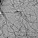
As seen from Table 1, ABM binding characteristics insignificantly differ for insulin and globin, suggesting that these proteins in fibrillar state have structurally similar ABM binding sites.
This work was supported by the grants from European Social Fund (project number 2009/0205/1DP/1.1.1.2.0/09/APIA/VIAA/152) and Fundamental Research State Fund of Ukraine (project number F.41.4/014).
Interaction of Fibrillar Insulin and Globin with Liposomes: Resonance Energy Transfer Study
M.Romanova*1, I.Maliyov1, A.Kastorna1, K.Vus1, V.Trusova1, J.Molotkovsky2, G.Gorbenko1
1V.N. Karazin Kharkov National University, Kharkov, Ukraine;
2Institute of Bioorganic Chemistry, Russian Academy of Sciences, Moscow, Russia
e-mail: romanova-mari@mail.ru
A wide range of proteins are capable of assembling into cross β-sheet structures (amyloid fibrils) under pathological or nonphysiological conditions. The toxicity of amyloid fibrils and their precursors is supposed to be associated with their action on structural and functional state of biological membranes. One way of approaching this problem is based on the use of model systems consisting of lipid vesicles and protein aggregates formed in vitro.
The present study was directed towards clarifying the molecular details of interaction between globin or insulin fibrils and liposomes of different composition. Fibrillar aggregates were obtained by protein incubation in glycine buffer (pH 2.2) at 60oC during 8 days. To assess the extent of fibril binding to liposomes, we measured energy transfer efficiency from tryptophan residues of globin or insulin to anthrylvinyl (AV) fluorophore covalently attached to phosphatidylcholine (PC). Lipid vesicles were prepared from PC and its mixtures with anionic lipid phosphatidylserine (PS) (10, 20, 40 mol% PS). Increasing the concentration of globin fibrils resulted in the raise of AV fluorescence intensity for all model systems under study (Fig.1). It was found that the most effective energy transfer occurs when globin interacts with neutral PC vesicles. In the case of insulin, conversely, the energy transfer from Trp to AV-PC was negligible. This observation can be explained by the fact that insulin has negative charge under the employed experimental conditions (pH 7.4), which prevents protein binding to negatively charged PC/PS liposomes.
|
|
|
|
Fig.1. Efficiency of energy transfer as a function of fibrillar globin concentration |
Fig.2. Comparison of energy transfer from Trp to AV-PC for insulin and globin |

Fig.2 illustrates the difference between fibrillar insulin and globin in the energy transfer efficiency from Trp to AV-PC in PC bilayers. The results obtained allowed us to suppose that the character of interaction between globin fibrils and liposomes is mostly hydrophobic, while in the case of insulin the role of electrostatics may be predominant. This work was supported by the grant from Fundamental Research State Fund of Ukraine (project number F.41.4/014).
Fluorescence Approaches to Detection of Protein Aggregates
V.M.Trusova
V.N. Karazin Kharkov National University, Kharkov, Ukraine
valtrusova@yahoo.com
Fluorescence spectroscopy is one of the most powerful tools for characterization of a multitude of biological processes1. Of these, the phenomenon of protein oligomerization attracts especial interest due to its crucial role in the formation of fibrillar protein aggregates (amyloid fibrils) involved in ethiology of so-called protein misfolding diseases. It is becoming increasingly substantiated that protein fibrillization in vivo can be initiated and modulated at membrane-water interface2. All steps of membrane-assisted fibrillogenesis, viz., protein adsorption onto lipid bilayer, structural transition of polypeptide chain into a highly aggregation-prone partially folded conformation, assembly of oligomeric nucleus from membrane-bound monomeric species and fiber elongation can be monitored with a mighty family of fluorescence-based techniques3-5. Little or no damage to the examined matter, requirement of material micromolar concentration, rather simple methodology, involvement of relatively inexpensive instrumentation, high sensitivity and specificity make fluorometry a broadly used research tool for studying the protein-protein interactions. Various kinds of fluorescence technique, namely steady-state and time-resolved fluorescence, fluorescence polarization and fluctuation spectroscopy, stopped-flow and laser-induced fluorescence provide the information about the structure, microenvironment and distribution of protein complexes1. The immense range of parameters measured for both intrinsic and extrinsic protein fluorophores includes fluorescence intensity, quantum yield, anisotropy and quenching, lifetime, Förster resonance energy transfer efficiency (FRET) efficiency, diffusion coefficients, to name just a few. In the present contribution the applications of fluorescence spectroscopy to monitoring protein oligomerization in a membrane environment are exemplified and some problems encountered in such kinds of studies are highlighted. More specifically, such aspects as fluorescence probing of protein-membrane binding, fluorescence monitoring of protein structural changes, detection of protein aggregates with intrinsic and extrinsic fluorophores are discussed. In addition, optimal strategies for fluorescence analysis of protein aggregation are suggested.
References:
1.Lakowicz, J.R. Principles of Fluorescence Spectroscopy. NY: Springer, 2006.
2.Gorbenko, G.P., Kinnunen, P.K.J. The Role of Protein-Lipid Interactions in Amyloid-Type Protein fibril Formation. Chem. Phys. Lipids, 2006, vol. 141, p. 72-82.
3.Zerovnik, E. Amyloid-fibril Formation. Proposed Mechanisms and Relevance to Conformational Disease, Eur. J. Biochem., 2002, vol. 269, p. 3362-3371.
4.Domanov, Ye.A., Kinnunen, P.K.J. Islet Amyloid Polypeptide Forms Rigid Lipid–Protein Amyloid Fibrils on Supported Phospholipid Bilayers. J. Mol. Biol., 2008, vol. 376, p. 42-54.
5.Trusova, V.M., Gorbenko, G.P., Sarkar, P. et al. Forster Resonance Energy Transfer Evidence for Lysozyme Oligomerization in Lipid Environment. J. Phys. Chem. B, 2010, vol. 114, p. 16773-16782.
Fluorescence Study of Cytochrome c Binding to Model Membranes
V.Trusova*1, G.Gorbenko1, J.Molotkovsky2, P.Kinnunen3
1V.N. Karazin Kharkov National University, Kharkov, Ukraine;
2Institute of Bioorganic Chemistry, Russian Academy of Sciences, Moscow, Russia;
3School of Science and Technology, Aalto University, Espoo, Finland
e-mail: valtrusova@yahoo.com
Cytochrome с (cyt c) is a small highly basic hemoprotein abundant in the intermembrane space of mitochondria and possessing two at first glance unrelated functions. First, cyt с is an essential component in respiration, shuttling electrons from cyt c reductase (bc1 complex) to cyt c oxidase. The second function of cyt c relates to its key role in the activation of the apoptotic cascade, resulting from the release of this protein from mitochondria into the cytosol. It is generally established that control of the binding of cyt c to the inner membrane of mitochondria (IMM) is involved in both of the above functions. Despite the diversity of lipids constituting the mitochondrial membrane, central to the cyt c – membrane interaction in IMM is cardiolipin (CL), which is indispensible for the retention, stability and physiological functioning of cyt c. In this regard, the nature and specificity of interactions between cyt c and CL attract particular interest. In the present study fluorescence resonance energy transfer (FRET) between anthrylvinyl-labeled phosphatidylcholine as a donor and heme moiety of cytochrome c (cyt c) as an acceptor has been employed to explore the protein binding to model membranes, composed of phosphatidylcholine (PC) and CL. The existence of two types of protein-lipid complexes has been hypothesized where either deprotonated or partially protonated CL molecules are responsible for cyt c attachment to bilayer surface. To quantitatively describe cyt c membrane binding, the adsorption model based on scaled particle and double layer theories has been employed, with potential-dependent association constants being treated as a function of acidic phospholipid mole fraction, degree of CL protonation, ionic strength and surface coverage. Multiple arrays of FRET data obtained under conditions of varying pH, ionic strength, CL content and protein-to-lipid molar ratio have been analyzed in terms of the model of energy transfer in two-dimensional systems combined with the adsorption model allowing for area exclusion and electrostatic effects. The set of recovered model parameters included effective protein charge, intrinsic association constants and heme distance from the bilayer midplane for both types of protein-lipid complexes. Upon increasing CL mole fraction from 10 to 20 mol % (the value close to that characteristic of IMM) the binding equilibrium dramatically shifted towards cyt c association with partially protonated CL species. The estimates of heme distance from bilayer center suggest shallow bilayer location of cyt c at physiological pH, while at pH below 6.0 the protein tends to insert into membrane core. These findings support the viewpoint that hydrogen bonding of cyt c to protonated phosphate can be coupled with the formation of electrostatic contacts with deprotonated phosphate. Structural features of cyt c do not exclude such a possibility. Cyt c molecule has two hydrophobic channels connecting heme crevice with the protein surface. The opening of one of these channels is surrounded by oppositely located Asn52 and Lys72, Lys73. It has been hypothesized that cyt c association with CL involves hydrogen bonding of one protonated phosphate moiety to Asn52 with simultaneous electrostatic binding of deprotonated phosphate to lysine residues and accommodation of one acyl chain within the hydrophobic channel.
This work was supported by the grants from European Social Fund (project number 2009/0205/1DP/1.1.1.2.0/09/APIA/VIAA/152) and Fundamental Research State Fund of Ukraine (project number F.41.4/014).
Aminobenzanthrone Dyes as Prospective Fluorophores for Detection and Characterization of Amyloid Fibrils
K.Vus*1, V.Trusova1, G.Gorbenko1, E.Kirilova2, G.Kirilov2, I.Kalnina2
1Department of Biological and Medical Physics, V. N. Karazin Kharkiv National University, Kharkiv, Ukraine;
2Department of Chemistry and Geography, Daugavpils University, Daugavpils, Latvia
e-mail: katenka.vus@mail.ru
In view of dramatic role of pathological protein aggregates, amyloid fibrils, in the development of a number of severe diseases, detection and characterization of such entities represent the focus of current research efforts. Fluorescence spectroscopy has been extensively employed in amyloid studies due to its inherent sensitivity and versatility, good spatial and temporal resolution. The aim of this work was to test a series of novel fluorescent aminobenzanthrone dyes (BD) for their ability to identify and characterize fibrillar aggregates of lysozyme prepared by protein denaturation in concentrated ethanol solution (FI) [1] or at strongly acidic pH (FII) [2]. Analysis of the results of fluorimetric titration in terms of the Langmuir adsorption model yielded the parameters of BD binding to native and fibrillar protein (association constant, binding stoichiometry and molar fluorescence). Furthermore, several additional quantities reflecting the preference of the probe to fibrillar protein aggregates have been evaluated [3]. Based on the comprehensive analysis of the recovered parameters, AM2 and A8 were selected as the most prospective tracers for FI and FII, respectively. Besides, A6 and A8 were also suitable for highly sensitive detection of both FI and FII types of amyloid fibrils, because of their high quantum yields in amyloid-bound state. Interestingly, comparison of quantum yields and binding parameters of the dyes in FI- and FII-bound states showed the differences in fibrillar morphologies. It was supposed that in FII fibers protofilaments are packed more tightly than in FI, that complicates BD penetration into the FII fibrils and is presumably responsible for the decrease of BD-protein binding extent. Finally, strong sensitivity of the novel probes to environmental viscosity was revealed by analyzing their properties in ethanol (E) and in ethanol/glycerol mixture with glycerol content of 67% (v/v) (EG). Substantially higher fluorescence anisotropy of BD in EG compared to E is indicative of strong viscosity sensitivity of the novel dyes. These findings suggest that fluorescence anisotropy of BD can be employed as an alternative parameter for studying the kinetics of amyloid fibril formation [4]. The present study demonstrated high potential of aminobenzanthrone dyes in identification of fibrillar protein aggregates and uncovering their structural peculiarities.
This work was supported by the grants from European Social Fund (project number 2009/0205/1DP/1.1.1.2.0/09/APIA/VIAA/152) and Fundamental Research State Fund of Ukraine (project number F.41.4/014).
References:
1. Holley, M., Eginton, C., Schaefer, D., Brown, L.R. Characterization of Amyloidogenesis of Hen Egg Lysozyme in Concentrated Ethanol Solution. Biochem Biophys Res Commun, 2008, vol. 373, p. 164-168.
2. Morozova-Roche, L.A., Zurdo, J., Spencer, A., Noppe, W., Receveur, V., Archer, D.B., Joniau, M., Dobson, C.M. Amyloid Fibril Formation and Seeding by Wild-Type Human Lysozyme and Its Disease-Related Mutational Variants. J Struct Biol, 2000, vol. 130, p. 339-351.
3. Vus, K., Trusova, V., Gorbenko, G., Kirilova, E., Kirilov, G., Kalnina, I., Kinnunen, P. Novel Aminobenzanthrone Dyes for Amyloid Fibril Detection. Chem Phys Lett, 2010, vol. 532, p. 110-115.
4. Sabate, R., Saupe, S.J. Thioflavin T Fluorescence Anisotropy: an Alternative Technique for the Study of Amyloid Aggregation. Biochem Biophys Res Commun, 2007, vol. 360, p. 135-138.
Binding of the Novel 4-tricyanovinylarylamine Dye to Fibrillar Lysozyme
K.Vus*1, V.Trusova1, G.Gorbenko1, T.Deligeorgiev2, S.Kaloyanova2, N.Lesev2
1Faculty of Physics and Technology, V. N. Karazin Kharkiv National University, Kharkiv, Ukraine;
2Faculty of Pharmacy and Chemistry, University of Sofia “St. Kliment Ohridsky”, Sofia, Bulgaria
e-mail: katenka.vus@mail.ru
Accumulation of
fibrillar protein aggregates (amyloid fibrils) in different tissues is
currently associated with a variety of so called conformational diseases. The
most widespread techniques employed in identification of amyloid fibrils are
fluorescence and absorption spectroscopy. Further progress in this research
area is strongly coupled with the design and testing of the novel chromo- and
fluorophores. The present study was aimed at evaluation of the novel 4-tricyanovinylarylamine dye (referred here
as 3q) as possible marker for fibrillar protein aggregates. Amyloid fibrils of
lysozyme were prepared at strongly acidic pH [1].
Tricyanovinylarylamine probes
are characterized by high extinction coefficients and the ability to form
intramolecular charge transfer state upon excitation[2]. Fluorescent
properties of these dyes are sensitive to their molecular structure and local
environment. It appeared that 3q has very low quantum yields in buffer solution
and there was no fluorescence increase in the presence of amyloid fibrils. For
this reason, we tested this dye as possible analog of the traditional amyloid
marker Congo Red, non-fluorescing dye whose differential absorption spectra are
used for selective detection of amyloid fibrils. As seen in Fig. 1A, increasing
the concentration of fibrillar lysozyme leads to progressive decrease of 3q
absorption, with clear isosbestic point. Quantitative characteristics of the
dye binding to protein aggregates (![]() – association constant, n –
binding stoichiometry, α – the difference of extinction
coefficients in the bound and free states) were estimated by analysing the
isotherm of 3q binding to lysozyme fibrils in terms of Langmuir
adsorption model (Fig. 1B). The recovered parameters are close to those
reported for existing amyloid-sensitive chromophores.
– association constant, n –
binding stoichiometry, α – the difference of extinction
coefficients in the bound and free states) were estimated by analysing the
isotherm of 3q binding to lysozyme fibrils in terms of Langmuir
adsorption model (Fig. 1B). The recovered parameters are close to those
reported for existing amyloid-sensitive chromophores.
|
A |
B |
|
Fig.1. Differential absorption spectra of 3q (A) and the isotherm of 3q binding to fibrillar lysozyme (B). |
|
To conclude, spectral behavior of 3q as one representative of tricyanovinylarylamine dyes allowed us to recommend these compounds for testing as possible amyloid markers similar to Congo Red.
This work was supported by the grants from European Social Fund (project number 2009/0205/1DP/1.1.1.2.0/09/APIA/VIAA/152) and Fundamental Research State Fund of Ukraine (project number F.41.4/014).
Association of Novel Benzanthrone Probes with Model Lipid Membranes
O.A.Zhitniakivska*1, V.M.Trusova1, G.P.Gorbenko1, E.M.Kirilova2, G.K.Kirilov2, I.Kalnina2
1V.N. Karazin Kharkiv National University, 4 Svobody Sq., Kharkiv, 61077, Ukraine; 2Department of Chemistry and Geography, Faculty of Natural Sciences and Mathematics, Daugavpils University, 13 Vienibas, Daugavpils LV5401, Latvia
e-mail: olya_zhitniakivska@yahoo.com
Among a wide variety of fluorescent dyes currently used in biomedical research and industry benzanthrone derivatives are of particular interest due to their favorable spectral properties, viz. large extinction coefficient, marked Stokes shift; negligible fluorescence in an aqueous phase; high sensitivity of fluorescence parameters to environmental polarity, etc. Our recent study revealed high lipid-associating ability of a series of newly synthesized benzanthrone aminoderivatives. Moreover, spectral responses of the two examined dyes in different lipid systems proved to correlate with increased bilayer hydration. These findings allowed us to assume that benzanthrone aminoderivatives may be effective fluorescent probes for examining membrane-related processes, especially those coupled with the change in the degree of lipid bilayer hydration. In view of this it seemed of interest to extend our previous investigation to a new series of benzanthrone dyes (referred to here as AM12, AM15, AM18, IAH, IBH and ISH), whose structures are given in Fig.1.
|
|
|
|
|
|
|
|
|
Fig. 1. Structures of aminobenzanthrones
|
||


The binding of aminobenzanthrones to the lipid membranes composed of zwitterionic lipid phosphatidylcholine (PC) and its mixtures with cholesterol (Chol) and anionic lipid cardiolipin (CL) was followed by significant increase in fluorescence intensity and dye quantum yields. To characterize benzanthrone-lipid binding quantitatively, we determined the dye partition coefficients for different lipid systems. CL and Chol addition to PC bilayer resulted in the increase of partition coefficients for IBH, ISH and IAH (relative to the neat PC membrane), whereas AM12, AM15 and AM18 displayed the opposite behavior in cardiolipin- and cholesterol-containing systems. As spectral properties of benzanthrone dyes strongly depend on the nature of fluorophore environment (polarity, viscosity, the probability of hydrogen bonding or other intermolecular interactions), it seems reasonable to interpret the differences between IBH, ISH, IAH and AM12, AM15, AM18 in terms of different bilayer location. It was assumed that ISH, IBH and IAH are located in the bilayer polar region, while AM12, AM15 and AM18 penetrate more deeply, most probably in the membrane hydrophobic part.
This work was supported by the grants from European Social Fund (project number 2009/0205/1DP/1.1.1.2.0/09/APIA/VIAA/152) and Fundamental Research State Fund of Ukraine (project number F.41.4/014).
New ICT-dyes for Membrane Studies
O.Zhytniakivska*1, V.Trusova1, G.Gorbenko1, T.Deligeorgiev2, S.Kaloyanova2, N.Lesev2
1V.N. Karazin Kharkiv National University, 4 Svobody Sq., Kharkiv, 61077, Ukraine;
2Faculty of Pharmacy and Chemistry, University of Sofia, Sofia, Bulgaria
e-mail: olya_zhitniakivska@yahoo.com
During the past decades fluorophores which can undergo photoinduced intramolecular charge transfer (ICT dyes) find increasing application in biological sciences. Fluorophores which exhibit ICT state are widely used in different areas, including the development of solar cells and chemosensing. High sensitivity of these compounds to the environmental polarity provokes their use as fluorescent microenvironmental sensors, particularly, in the studies of membrane structure and protein-lipid interactions.
The present study was undertaken to evaluate the sensitivity of newly synthesized ICT dyes, referred to here as ICT2, ICT4, ICT3 and ICT5, whose structures are given in Fig.1, to the changes in physicochemical properties of lipid bilayer. At the first step of the study we compared the lipophilic properties of the ICT-dyes and their sensitivity to the membrane environment. Fluorescence spectra of these dyes were recorded in buffer solution (5 mM Na-phosphate, pH 7.4) and liposomal suspension. It appeared that only ICT2 and ICT4 are capable of fluorescing, while ICT3 and ICT5 are non-fluorescent. ICT4 fluorescence decreases, while emission maximum is shifted from 435 nm in buffer to about 420 nm on the dye transfer from the aqueous to lipid phase. On the contrary, the binding of ICT2 to the model membranes was followed by the fluorescence increase without any shift of the emission maximum. Moreover, fluorescence quantum yield of ICT2 exhibited tenfold increase in the lipid bilayer compared to the buffer solution, while that of ICT4 decreased about twice.
|
|
|
|
|
|
|
Fig. 1. Structure of the dyes ICT2 (A), ICT4 (B), ICT3(C) and ICT5 (D) |
|


To characterize
ICT2-lipid binding quantitatively, at the next step of the study we determined
the dye partition coefficient ![]() for different lipid systems. The values
of
for different lipid systems. The values
of ![]() were
found to fall in the range (0.4 – 1.5)×104 indicating that the
examined dyes possess high lipid-associating ability. Analysis of the partition
coefficients shows that inclusion of anionic cardiolipin and cholesterol into
phosphatidylcholine bilayer gives rise to the increase of partition coefficient
relative to the neat phosphatidylcholine membrane.
were
found to fall in the range (0.4 – 1.5)×104 indicating that the
examined dyes possess high lipid-associating ability. Analysis of the partition
coefficients shows that inclusion of anionic cardiolipin and cholesterol into
phosphatidylcholine bilayer gives rise to the increase of partition coefficient
relative to the neat phosphatidylcholine membrane.
To summarize, a comparison of the fluorescence properties of the four newly synthesized ICT compounds revealed that despite being structurally similar, these dyes display different spectral and lipid-associating behavior. ICT2 was featured by the most effective partitioning into lipid phase coupled with considerable increase of the fluorescence quantum yield. The recovered properties of ICT2 allowed us to recommend this dye for further use in membrane studies. This work was supported by the grant from Fundamental Research State Fund of Ukraine (project number F.41.4/014).
Alterations of Blood Plasma Albumin in Chernobyl Clean-up Workers with and without Type 2 Diabetes Mellitus
T.Zvagule2, N.Kurjane2, A. Šķesters2, A.Silova2, N.Gabruseva2, G.Gorbenko3, G. Kirilov1, I.Kalnina1, E.Kirilova1, S.Gonta4
1Daugavpils University, Daugavpils,
2Riga Stradina University, Riga, Latvia,
3V.N. Karazin National University, Kharkov, Ukraine;
4Institute of Microbiology and Biotechnology, Latvia University
Diabetes mellitus has been one of the most crippling diseases that man has seen and its prevalence has risen dramatically over the past two decades. Currently there are over the 150 million diabetics word wide and this number is likely to increase to 300 million or more by the year 2025. Diabetes mellitus increases the risk of many disorders including cardio-vascular diseases. Understanding the molecular properties of diabetic progression is a big challenge in systems-biology era.
ABM (3-aminobenzanthrone derivative) has been previously shown as a potential biomarker for determination of the immune state of patients with different pathologies. The aim of this study was to determine the several aspects of plasma albumin alterations in the group of Chernobyl clean-up workers with diabetes mellitus in relation with its common group without diabetes mellitus and humans having no professional contact with radioactivity. Patients: 1 Group type 2 diabetes mellitus. Complications associated with diabetes- such as polyneuropa5thy, atherosclerosis, including chronic diseases with inflammation. Group 2 – prevalence of several chronic diseases (diabetes mellitus is as concomitant disease. Cardiovascular disorders, progressive atherosclerosis with active inflammation process, leucocytosis, metabolic syndrome, stroke. Group 3 – Chernobyl clean-up workers without diabetes mellitus. ABM fluorescent characteristics are dependent on functional activity of CNS and indices of inflammatory processes. The most serious combination for patients-holisterinemy, progressive cerebral atherosclerosis, epilepsy paroxysms. The following parameters were examined: 1) the spectral characteristics of ABM in blood plasma; 2) “’effective” (EA) and total (TA) albumin concentration in blood plasma; 3) quantitative parameters of albumin auto-fluorescence.
Screening of the individuals with diabetes mellitus 25-26 years after the work in Chernobyl revealed two groups of patients differing in structural/functional properties of albumin, first of all on conformations of plasma albumin and characteristics of tryptophanyl region of molecule. The revealed structural modifications of membranes are dependent on radiation- induced factors. Concomitant diseases (diabetes mellitus, cardio-vascular diseases) reinforce radiation induced effects in accordance with its manifestation. Results clarifies the heterogenous nature of albumin molecules and revealed quantitative different its conformation in observed groups of patients. Albumin molecule in diabetics is modified in the chronic hypoxia conditions provoked mainly by the hyperglycemia and oxidative stress.
ABM is a sensitive probe of albumin alterations, and can be used to elucidate the changes in protein systems. Significant differences in albumin dynamics exist between control (donors), and diabetics and non-diabetics groups of Chernobyl clean-up workers. The biomarker of modified albumin could also help to identify individuals with complex metabolic conditions who have a higher risk of suffering from cardiovascular diseases.
Alterations of Blood Plasma Albumin in Chernobyl Clean-up Workers with and without Concomitant Diseases
T.Zvagule1, I.Kalnina2, E.Kirilova2, N.Kurjane1, N.Gabruseva1, J.Reste1, G.Kirilov2,
A. Šķesters1, A.Silova1
1Riga Stradins University, Latvia
2Daugavpils University, Daugavpils, Latvia
ABM (3-aminobenzanthrone derivative) developed at Daugavpils University has been previously shown as a potential probe for the determination of the immune state of patients with different pathologies. It is widely accepted that the dynamics of plasma proteins (e.g. albumin) play a prime role immune characteristics of humans. Albumin is a single source of ABM fluorescence in human blood plasma (650 nm). The aim was to determine the several aspects of blood plasma albumin alterations of Chernobyl clean-up workers, who had worked within the zone from 1986 till 1991. Most of them were officially documented as recipients of ionizing radiation exposure (1-50 cGy). For the study were selected the different groups of Chernobyl clean-up workers groups (common, after myocardial infarction, with epilepsy paroxysm, and 2 type diabetes mellitus), and corresponding groups of patients having no professional contact with radioactivity. The following parameters were examined: 1) the spectral characteristics of ABM in blood plasma, 2) plasma auto-fluorescence parameters; 3) “effective” and total albumin concentrations in blood plasma. (“Effective „concentration –equivalent in plasma of healthy albumin and reflects its binding and carrier functions). The total albumin concentration is more conservative. It has been noted that changes of pH in range from 3-12 strongly affect spectral characteristics of albumin bound ABM. The spectral characteristics of ABM in Chernobyl clean-up workers plasma results in splitting of the albumin alterations onto two stages, acidic expansion stage (620-630 nm) and N-F transition stage (600-620 nm).These observations may be consistent with the decreased binding of ABM and/or conformational changes of albumin molecule, significantly differing in observed patients groups. The levels of pathological and pharmacological metabolites (fatty acids, antioxidants, plasma levels of lipid peroxidation products etc.) balance differs in patients groups comparable to controls and hence their correlation to seizures pathophysiology and their degree. The metabolites caused conformational changes in albumin molecule and shifts in binding parameters are in agreement with results of albumin auto-fluorescence data and ABM binding sites characteristics (binding sites count, affinity for probe, polarity). A result clarifies the heterogeneous nature of ABM binding and revelled quantitative different conformation of albumin in observed groups of patients. The spectral parameters of ABM in plasma (as in lymphocytes) are coupled with cellular mechanisms of immune regulation in patients. The results showed strong correlation with select immunological parameters (CD4+: CD8+ ratios, lymphocyte count etc.) and both clinical and pathological estimates. The more pronounced albumin structural/functional alterations were observed in clean-up workers with concomitant diseases. One can with a certain degree of confidence that the revealed alterations of albumin are dependent on radiation induced factors. Concomitant diseases reinforce radiation – induced effects in accordance of its manifestation. Therefore seems likely that external radiation and incorporated radionuclides) predominate in alterations of albumin. Measures of ABM characteristics in blood plasma could potentially be a useful tool in clinical immunological screenings to estimate the immune state of patients.
Aminobenzanthrone Derivative ABM - Fluorescent Biomarker of Immune State of Patients
E.Kirilova1, G.Gorbenko3, I.Kalnina1, T.Zvagule2, G.Kirilov1, N.Kurjane2
1Daugavpils University, Latvia;
2Riga Stradins University, Latvia;
3V.N.Karazin Kharkov National University, Kharkov, Ukraine
In different pathologies membrane damage in immune cells and blood plasma albumin envolves as a consequence of alterations in a immune state of patients. It is now widely accepted that the dynamics of these changes, along with the types of alterations in structures of the immune system cells and plasma proteins play a critical role in the maintenance of the immune status of any given organism. As a result of the importance of changes in the structural integrity of cells of the immune system, it is important for clinicians to receive information on the immunological status of organism via quick, reliable, reproducible methods. In this regard, fluorescent probes have shown to be excellent tools for use in such protocols. Such an analysis has a great potential for not only for helping to comprehed mechanisms of immunomodulation associated with the induction/progression of pathologies, but might serve as a very important prognostic indicator of long- term survival among patients with pathologies. The advantages of probe ABM:
1. Spectral characteristics of aminobenzanthrones (e.g. ABM) satisfy all requirements for an ideal tracer (bright fluorescence, high extinction coefficient, photo-, thermo-and chemical stability). Specifically, it was found that spectral characteristics of this probe correlate with a number of important parameters of artificial and cellular membranes such as physicochemical state, microviscosity, proliferating and lipid metabolic activities of cells, distribution of lymphoid subset, etc.
2. The choise to examined albumin, among the myriad of constituents in plasma, is that this protein is practically the single source of ABM binding and subsequent fluorescence in plasma. Very important is that ABM can be used as a probe sensitive to conformation changes in protein: the most prominent changes in fluorescence characteristics occured at pH values 3-12 known to cause conformation transitions of proteins. It is significant to note that the probe emission in red region of spectra (650 nm) exceeds higher analytical sensitivity of method (as compared with probes ANS, K-35, PR etc.). For example- according the literature data probe ANS possess low sensitivity to conformative changes of albumin at pH 5.5-11.5. Only fluorescent probes allow to detect the „effective” concentration of albumin in blood plasma („effective” concentration- equivalent in blood plasma of healthy albumin and reflects it’s binding and carrier functions). The total albumin concentration is more conservative. The detection of „effective ‘concentration of albumin using ABM is technically simple and not so time –consuming as in the cases of other probes (don’t require additional steps of plasma and probe preparation). ABM spectral parameters in blood plasma are coupled with alterations in cellular mechanisms of immune regulation in the patients with different pathologies. Measures of ABM fluorescence intensity values for plasma albumin and/or for lymphocytes (total and among different subtypes) could potentially be a useful tool in clinical immunilogical screenings to estimate the immune state of patients.
Functional State of Beta Adrenoreactive System in Man and Animals: Fluorescent Studies
E.Kirilova1, I. Kalnina1, T.Zvagule2, N. Kurjane2, G. Kirilovs1, N. Gabruseva2
1Daugavpils University, Daugavpils, Latvia,
2Riga Stradins University, Latvia
Analysis of the structural /functional organization cell membranes, specifically, the studies of the functional state of beta adrenoreceptors is a topical problem in medicine and biology. The direct detection of functional activity of adrenoreceptors is methodologically complete and therefore not suitable for systemic determination. Earlier works have shown the unidirectivity of the changes in the adrenoreceptive properties of various tissues and organisms (red blood cells including) during the pharmacological action on the adrenergic mechanisms of regulation in an organism. Thus, red blood cells are a convenient test object which makes it possible to judge on the changes in the adrenoreactivity of an organism on the whole.
We investigated the binding of n-toluene sulfonate-4-(n-dimethylaminostyrene)-I-methylpyridinium (DSM) to human and animal erythrocyte membrane. Probe –cation DCM possess such properties as polychromaticity, sensitivity to the changes in polarity, viscosity of medium. The binding of probe to the erythrocyte membrane was studied in pathological states (myocardial infarction, instable stenocardia etc.; at high and low adrenoreactivity of animals (quinea pigs, rats). Increased adrenoreactivity was due to intraperitoneal injection of quanethidine (30-50 mg/kg, 24 h); decreased adrenoreactivity by intraperitoneal injection of propranolol (5mg/kg, 20min).The emission spectra of DCM in erythrocytes consists of two bands with the maxima at 560-565 nm( exc. 470 nm) and 610-620 nm (exc. 495 nm). The differences in the DCM binding parameters to erythrocytes were found at low and high adrenoreactivity of animals and observed patients groups as compared with control groups. Lower binding constants K and higher number of binding sites N (an indication of less specific interactions) we found for the observed groups of patients as compared with healthy subjects. At an increased adrenoreactivity of the organism it was found that the values of N do up and those of K down. At the decreased adrenoreactivity there were lowering of N and rise of K. Changes of fluorescence intensity (610-620 nm) correlates with dynamics of change in parameters N and fluorescence intensity (560-565 nm). Similarity of the character of membrane response to the change in the adrenoreactivity of the organism during the effect of stress factors and in diseases of the cardio-vascular system suggest that the along structural rearrangement of erythrocytes membrane the structural and functional properties of the adrenoreceptors also change. Increased adrenoreactivity of animals led to enhanced affinity of erythrocytes beta adrenoreceptors to propranolol and their binding to fluorescent robe were changed. It is concluded that probe DSM are promising in studies of the changes in membrane properties and in the beta-adrenoreceptive function both the erythrocytes and the organism as a whole.
Fluorescent Studies of Human Blood Plasma Albumin Alterations and Lymphocytes Subpopulations in Colorectal Cancer Patients
I. Kalnina1, T. Zvagule2, L. Klimkane3, N. Kurjane3, E. Kirilova1, G. Gorbenko1
1 Daugavpils University, Latvia;
2 Riga Stradins University, Latvia;
3 Latvian Centre of Oncology, Riga, Latvia;
The novel fluorescent probe ABM was used to characterize lymphocyte membranes and blood plasma albumin from colorectal cancer patients in context of the hosts immunological parameters and state of cancer progression. For study patients with colorectal cancer were examined: 10 one day before and 10 days after their surgical treatment ; 2) as disease worsened (Stages IIa, IIIb, IV); 3) advanced cancer patients. The aim of studies was to evaluate the potential utility of measures of ABM fluorescence parameters as a standart tool in the analysis of host immune status and for a clinical interpretation of alterations in albumin per se and lymphocytes functional activity in patients. ABM spectral parameters in lymphocytes reflects interrelated properties of the cells: 1) outer membrane physicochemical state, 2 )membrane microviscosity, 3) proliferative activity, 4) lipid metabolism and/or 5) phenotypical profile. ABM binds with blood plasma albumin with high level of selectivity. Probe ABM is very sensitive to all known conformational changes of albumin in the region pH 3-12. Spectral parameters of ABM binding with plasma albumin reflect albumin "effective" concentration (equivalent of "healthy" albumin in patients plasma), alterations of albumin globule, its physical and functional properties, characteristics of binding sites properties. The significant decrease of ABM fluorescence intensity in plasma could be explained by binding capacity/conformational changes of the albumin in these patients. Specific relationship was obtained between ABM fluorescence in lymphocytes and characteristics of cells: anisotropy index, binding constant, binding sites number etc. The lymphocyte distribution among the subsets also differed. Interestingly, the ABM fluorescence intensity in the cell suspension and blood plasma correlate with select immunological parameters (CD4+, CD8+, ratio CD4+/CD8+, CD38+, CD16+ , level of immunoglobulines IgA, IgG, IgM etc.). Decrease in the CD4+/CD8+ ratio mainly depend on an increase in the T- suppressor cells in patients without metastases, whereas it is due to a decrease in the T-helper cell in most patients with metastatic disease. Surgical treatment affects immunological parameters and apperead to elevate lymphocytes functional activity. The preoperative immune state of patients is important for their survival. Immunosupression increased gradually with progress of cancer; capacity of albumin binding reserve and „effective” concentration decrease. These findings suggest likely physical (structural) and functional alterations in the patients cells and plasma were a function of cancer stage. In advanced cancer, in contrast to other groups, the absolute number of lymphocytes had direct (not inverse) correlation with ABM fluorescence intensity.The higher level of lymphocytes number, T-cell proliferative activity, and albumin ‘’effective ‘’ concentration have a beneficial effect on overall survival. There was excellent agreement between changes in spectral characteristics and both clinical and pathological estimates of disease severity. Measures of ABM fluorescence intensity values in blood plasma and lymphocytes (as reflection of their functional activity) might be a useful tool in the evoluation of the immune status of patients in clinics, including prognosis, prediction of therapeutic efficacy, treatment outcomes. The fluorescence-based method is less expensive, not very time-consuming, technically simple, 100 times more sensitive than standart ones.
Oksidatīvā stresa marķieru izmaiņas Černobiļas AES avārijas seku likvidētājiem pēdējo 10 gadu laikā
A. Šķesters1, A. Silova1 , T. Zvagule2, J. Reste2, L. Lārmane1, Ņ. Rusakova1, N. Gabruševa2.
1 RSU Bioķīmijas laboratorija, Rīga, Latvija;
2 Paula Stradiņa KUS Aroda un radiācijas medicīnas centrs, Rīga, Latvija
1986. gada 26. aprīlis ir kļuvis par neatgriezenisku likteņa pavērsiena punktu vairāk kā 6000 tūkstošiem Latvijas iedzīvotāju un viņu ģimeņu. Šī diena mainīja ne tikai cilvēku dzīves, bet ienesa ievērojamas izmaiņas visas Eiropas saimnieciskajā un finanšu sistēmā.
Padomju „ kodolzinātnes eksperimenta” rezultātā, uz cilvēkiem iedarbojās ne tikai jonizējošā radiācija, bet atmosfērā nonāca radioaktīvie izotopi ( 137Cs, 90Sr, Ru, 131J) un putekļi, cilvēki cieta no nekvalitatīvas pārtikas, ūdens, fiziskas pārpūles un psiho-emocionālās spriedzes. Šo kaitīgo faktoru rezultātā, jau sākotnēji konstatēja akūtu oksidatīvo stresu, kurš vēlāk pārgāja hroniskā formā. Pašreiz, attiecībā uz Černobiļas AES avārijas seku likvidētājiem, pieņemts lietot terminu – ilgstošs hronisks oksidatīvais stress, kura, pastāvīgas un ilgstošas iedarbības laukā, ir lipīdi un proteīni, kuri lokalizēti šūnu membrānas intra- un ekstra-celulārās struktūras, gan arī enzīmi un hormoni. Oksidatīvo stresu un tā pakāpi raksturojošos marķierus var iedalīt pēc to veidošanās – no lipīdu oksidācijas procesa (LPO) nākušie, un tie marķieri, kuri veidojas proteīnu oksidācijas (PO) laikā. Bez tam, oksidatīvā stresa lielumu raksturo enzīmi-antioksidanti: Cu,Zn-superoksīddismutāze, katalāze, glutationa peroksidāze, tādi ne-enzimatiskie antioksidanti, kā vitamīns E, albumīni, selēns, arī varš, cinks, askorbāts, un citi. Mēs salīdzinājām dažus no minētajiem oksidatīvā stresa marķieriem, kurus analizējām I posmā (1998/99. gadā) un II posmā (2010/11. Gadā).
Nosakot lipīdu oksidācijas intensitāti I posma laikā, konstatējām ievērojamu oksidācijas procesu intensificēšanos – lipīdu hidroperoksīdu (LOOH) saturs asins plazmā trīskārtīgi pārsniedza LOOH saturu praktiski veselu atbilstoša vecuma vīriešu asinīs. Gandrīz dubultīgi bija pārsniegta lipīdu (nepiesātināto taukskābju) oksidējamība. LPO procesa norises ātrums ČAES avārijas seku likvidētājiem, vairāk kā 3 reizes pārsniedza normu. Ievērojami palielināts izrādījās arī malondialdehīds (MDA). Tai pašā laikā, avārijas seku likvidētāju asinīs, konstatējām ievērojamu selēna deficītu, kurš svārstījās robežās no neliela ( 68 ± 10 µg/L ) līdz dziļam (46 ± 10 µg/L ) deficītam. Ievērojami samazināta bija glutationperoksidāzes aktivitāte, vitamīna E (α-tokoferola) saturs un kopējā antioksidatīvā organisma aizsardzība (AOA) asinīs. 10 gadus vēlāk, nosakot oksidatīvā stresa marķierus ČAES avārijas seku likvidētājiem (pirms antioksidantu un nesteroīdo pretiekaisuma līdzekļu indicēšanas), konstatējām, ka, gandrīz visi iepriekšējie marķieri saglabājuši savas izmaiņas attiecībā pret normu. Tiesa, daži no marķieriem ir ievērojami samazinājušies un tuvojas normas robežai (AOA, selēna un E vitamīna saturs asinīs), citi palikuši gandrīz nemainīgi (LOOH, MDA, lipīdu oksidējamība un LPO norises ātrums). Praktiski nemainīga saglabājusies kopējā organisma antioksidatīvā aizsardzība. Tādējādi apstiprinājās pieņēmums, ka ČAES avārijas seku likvidētāju organismā joprojām saglabājas hronisks oksidatīvais stress.
Pēc dabīgo antioksidantu (AO), ne-steroīdo pretiekaisuma (NSAID) līdzekļu vai AO/NSAID kombinācijas lietošanas, ČAES avārijas seku likvidētājiem, ievērojami samazinājās oksidatīvo stresu raksturojošie marķieri, daži – MDA, selēns, vitamīns E sasniedza zemāko normas robežu.
Tādējādi esam ieguvuši apstiprinājumu, ka dabīgos antioksidantus vēlams iekļaut kompleksajā terapijā gadījumos, kad oksidatīvā stresa marķieri uzrāda ievērojamas atšķirības no normas. Paralēlajā klīniskajā pētījuma daļā atklātas labvēlīgas izmaiņas klīniskajā ainā pēc dabīgo antioksidantu lietošanas. Tai skaitā: samazinājās pieprasījums pēc recepšu medikamentiem, kritās holesterīna līmenis asinīs, mazinājās locītavu un citas lokalizācijas sāpes, migrēna, depresija, kuņģa problēmas.
Jaunu - liposomas veidojošu katjonu karbazola atvasinājumu izmantošana šūnu transfekcijā in vitro
A. Vežāne1, G. Apsīte1, A. Baran2, R. Brūvere1, V. Ose2, A. Plotniece2, I. Timofējeva1, T. Kozlovska1
1 Latvijas Biomedicīnas pētījumu un studiju centrs (BMC), Rīga, Latvija
2 Latvijas Organiskās sintēzes institūts (OSI), Rīga, Latvija
e-pasts: aleksandra.vezane@biomed.lu.lv
Pēdējā laikā strauji pieaug interese par fluorescento krāsvielu – t.s. zonžu pielietojumu bioloģijā un medicīnā. Arī Latvijā, Latvijas Organiskas sintēzes institūtā (OSI), tiek sintezēti jauni bioloģiski aktīvi savienojumi, kuru fluorescentās īpašības tiek plaši pētītas Latvijas Biomedicīnas pētījumu un studiju centrā (BMC).
Konkrētā pētījuma mērķis bija izvērtēt 3 jauno - liposomas veidojošo katjonu karbazola atvasinājumu izmantošanas potenciālu, pielietojot tos šūnu transfekcijā in vitro.
Jauno - liposomas veidojošo katjonu karbazola atvasinājumu spēju veidot kompleksus ar ģenētisko materiālu (plazmīdas DNS), vērtēja, izmantojot gan elektronmikroskopiju, gan retardāciju agarozas gēlā. Jauno savienojumu spēja piegādāt ģenētisko materiālu kultivējamām eikariotu šūnām jeb transfekcijas efektivitāte – tika noteikta izmantojot fluorescento mikroskopu.
Ar tā palīdzību tika izvērtēts (GFP - zaļi fluorescējoša proteīna), ekspresijas līmenis, kuru ieguvām, ievadot šūnās plazmīdu pEGFP. Pētāmās šūnās tika transficētas arī ar plazmīdu pLac-Z. Šī fermenta β-galaktozidāzes aktivitātes pakāpe tika noteikta pielietojot spektrofotometriju. Lai noteiktu ekspresējošā GFP proteīna lokalizācijas vietu transficētās šūnās, tika pielietota konfokālā lāzera skenējošā mikroskopija.
Pētījumu rezultātā tika pierādīts, ka bioloģiski aktīvās fluorescentās zondes spēj ne tikai specifiski saistīties ar šūnu membrānas struktūrām, tā iezīmējot šūnas un padarot tās fluorescējošas, bet arī spēj saistīt ģenētisko materiālu, veidojot liposomas un tādējādi to transportēt mērķa šūnās. Šis darbs tika finansiāli atbalstīts no Valsts pētījumu programmas līdzekļiem.
- Chang, C.-C. Chemistry and Biodiversity, 1, 1377 (2004).
- Saengkhae, C. Eur. J. Pharm., 559, 124 (2007).
- Tanious, F. Biochemistry, 39, 12091 (2000).
- Dias, N. Biochemistry, 43, 15169 (2004).
- Chang, C.-C. J. Chin. Chem. Soc. 50, 185 (2003).
Glikogēna noteikšana raugos S. cerevisiae izmantojot jaunus uz benzantrona bāzes sintezētos fluoroforus
I.Kralliša
Latvijas Universitātes Mikrobioloģijas un biotehnoloģijas institūts, Rīga, Latvija
krallish@latnet.lv
Fluorescentās metodes mūsdienās uzskata par progresīvu un efektīvu pieeju mikroorganismu fizioloģijas pētījumiem, kā arī biotehnoloģijas jomā. Izmantojot uz benzantrona bāzes sintezētās fluorescentās zondes, tika veikti Saccharomyces cerevisiae iekš šūnu rezerves metabolīta glikogēna biosintēzes pētījumi oksidatīvā stresa - 10% (v/v) spirta barotnē un normālos augšanas apstākļos. S. cerevisiae ir vērtīgo bioloģiski aktīvo vielu producents un plaši izmantots pētījumos kā eukariotisko šūnu modelis, tāpēc tas ir ļoti perspektīvs un piemērots modelis fluorescento metožu izstrādei ar mērķi pētīt eikariotisko organismu šūnu noturības mehānismus dažādu stresa faktoru ietekmei uz šūnu fizioloģiju.
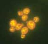
Lai atlasītu efektīvas fluorescentās zondes tika pārbaudīta jauniegūto zonžu piemērotība un aktivitāte attiecībā pret mērķa savienojumu glikogēnu, kurš tika izdalīts no S. cerevisiae šūnām. Fluorescento zonžu efektivitāte tika salīdzināta ar komerciālo glikogēnam specifisko akriflavīna efektivitāti. Kā efektivitātes rādītāji tika ņemti zonžu spektrālu parametru (emisijas maksimums un fluorescences intensitāte) atkarība no glikogēna koncentrācijas. Tika pārbaudītas vairākas fluorescentās zondes izmantojot glikogēna standarta šķīdumu , kā arī glikogēns tika izdalīts no S. cerevisiae šūnām augot sintētiskajā Ridera barotne vai YPG barotnē. Glikogēns tika izdalīts no raugu šūnām izmantojot 0,25 M Na2CO3 (sārmainā frakcija) un 0,5M HClO4 (skābā frakcija), kā arī dots kopējais glikogēna saturs šūnās. Dati iegūtie glikogēna satura noteikšanā, izmantojot akriflavīnu un atlasītos uz benzantrona bāzes sintezētos fluorescentos zondus tika salīdzināti ar glikogēna noteikšanu šūnu ekstraktos ar vispārpieņemto ķīmisko antrona metodi.
Pētījumu rezultātā tika parādīta pozitīva korelācija starp glikogēna saturu un fluorescences intensitāti izmantotos paraugos, kas ļauj izmantot jaunos uz benzantrona bāzes sintezētos fluorescentos zondus glikogēna satura ātram skrīningam raugu šūnu ekstraktos. Raugu šūnas oksidatīva stresa apstākļos palielina rezerves savienojuma glikogēna sintēzi, līdzīgi kā agrāk novērotam trehalozes uzkrājumam palielināšanai līdzīgos oksidatīva stresa apstākļos.
Alkilrezorcinolu izdalīšana un noteikšana baktērijās Azotobacter chroococcum.
R.Linde
Latvijas Universitātes Mikrobioloģijas un biotehnoloģijas institūts, Rīga, Latvija
Raimonds.linde@lu.lv
Veikta alkilrezorcinolu izdalīšana no baktērijām Azotobacter chroococcum ar Fast Blue B reaģentu un to noteikšana ar dažādām metodēm. Tika pārbaudīts alkilrezorcinolu daudzums baktērijās Azotobacter chroococcum, kas fermentētas dažādos vides apstākļos izmantojot spektrofotometru Shimadzu UV-260, kā arī ar plānslāņa hromatogrāfiju. Alkilrezorcinolu absorbcija tika noteikta ar vairākām standartvielām.
pH izmaiņu ietekme uz fluorescento krāsvielu emisijas spektriem
E.Pīmane
Latvijas Universitātes Mikrobioloģijas un biotehnoloģijas institūts, Rīga, Latvija;
epimane@inbox.lv
Pētījumam tika izmantotas vairākas jaunas benzantronu atvasinājumu krāsvielas. Eksperimentā tika pārbaudīta krāsvielu fluorescences īpašību noturība, mainoties vides pH līmenim. pH izmaiņas ietekmē krāsvielu fluorescences spektru emisijas maksimumus. pH līmeņi destilētam ūdenim tika regulēti ar 1N un 4N HCl un 0,1N, 1N un 4N NaOH. Mērījumi tika veikti, izmantojot fluorescences spektrofotometru Fluoromax3.
Fluorescentās krāsvielas P8 izmantošana polihidroksibutirāta un polihidroksibutirāta-ko-hidroksivalerāta lateksu kompozītplēvju raksturošanai
L. Savenkova
Latvijas Universitātes Mikrobioloģijas un biotehnoloģijas institūts, Rīga, Latvija;
azot@latnet.lv
No PHB un PHB/HV lateksiem tika izveidotas kompozītplēves, atšķirīgas pēc PHA granulu, polivinilspirta, glicerīna un fluorescentās krāsas P8 satura. Iegūtām PHA lateksa polimērplēvēm ir noteiktas mehāniskās īpašības. Ir secināts, ka kompozītplēvju sastāvs ļoti lielā mērā ietekmē gan mehāniskās īpašības, gan stiprību, gan elastību.
PHA lateksa kompozītplēvju virsmas struktūra tika vizualizēta ar konfokālā lāzera mikroskopijas metodi. Izveidoto kompozītplēvju izturību ūdens šķīdumos novērtēja pēc fluorescentās krāsvielas migrāciju inkubācijas vidē. Iegūtie dati par kompozītplēvju virsmas struktūras izmaiņām un fluorescentās krāsvielas migrāciju ļauj labāk izprast, kā lateksu kompozītu sastāvs ietekmē plēvju mehāniskās un fizikālās īpašības.
Dzelzs jonu ietekme uz izmaiņām šūnā, alkilrezorcīnu un PHB veidošanos Azotobacter chroococcum šūnās
E.Pīmane
Latvijas Universitātes Mikrobioloģijas un biotehnoloģijas institūts, Rīga, Latvija;
epimane@inbox.lv
Dzelzs jonu saturošiem savienojumiem ir ļoti liela nozīme atmosfēras slāpekli fiksējošai baktērijai Azotobacter chroococcum. Dzelzs daudzums ietekmē A.chroococcum augšanu un poli-3-hidroksibutirāta (PHB) veidošanos, kas ir oglekļa un enerģijas rezerves savienojums. A.chroococcum šūnas novecojoties vai arī stresa apstākļos no aktīvi dalošās veģetatīvās formas pāriet miera stāvoklī un pārvēršas par cistām. Cistu veidošanās ir liela nozīme alkilrezorcīniem, kas aizsargā šūnu no ārējās vides iedarbības. Vairāki pierādījumi liek domāt, ka metāli, tāpat kā alkilrezorcīni, var ietekmēt membrānu plūstamību. Fluorescentās metodes ir pietiekami jutīgas, ātras un tehniski vienkāršas un tādēļ var tikt izmantotas sākotnējai šūnu pārbaudei, lai noteiktu radušās izmaiņas.
Ar fluorescentajām un spektrofotometriskajām metodēm tika pārbaudīta dzelzs ietekme uz alkilrezorcīnu un PHB saturu šūnā. Izmaiņas šūnu suspensijas fluorescences intensitātē tika pārbaudītas ar fluorescento metodi, kur tika izmantota jauna, hidrofobā krāsviela P8 (3-piperidinobenzantrons).
Fluorescentie benzantrona atvasinājumi: kvantu-ķīmiskie aprēķini
S. Gonta
Latvijas Universitātes Mikrobioloģijas un biotehnoloģijas institūts, Rīga, Latvija;
svetago@lu.lv
Luminiscences krāsvielas ir atradušas plašu pielietojumu ķīmiskos un bioloģiskos pētījumos. Bet interese par jaunām fluorescējošam krāsvielām turpina pieaugt. Ir zināms, ka aizvietoti N-aril amidīni parāda intensīvu luminiscenci šķīdumā. Lai izpētītu elektroniskās struktūras un īpašības sintezētiem N-aizvietotiem amidīnu benzo[de]antracen-7-ons atvasinājumiem, tika veikti kvantu-ķīmiskie aprēķini.
Katram savienojumam veikta ģeometrijas optimizācija ar AM1 metodi. Aprēķini liecina, ka aromātiskam kodolam ir plakana konfigurācija, kā arī ir divi iespējamie virzieni no amidīna aizvietotāja. Aprēķina rezultāti korelē arī ar sarkano nobīdi savienojumu absorbcijas un emisijas spektros.
Amidīna aizvietotāji nerāda lielu ietekmi uz HOMO un LUMO enerģijām, kas aprēķinātas ar jebkuru no izmantotajām metodēm. Aizvietotāji galvenokārt ietekmē oglekļa atoma amidīna grupas, no otras puses, pie amidīna fragmenta esošie aizvietotāji ievērojami ietekmē molekulas dipolu momentu. Tas aug, ja pie amidīna fragmentu molekulas ir elektronu donoru grupas (Me, Et).
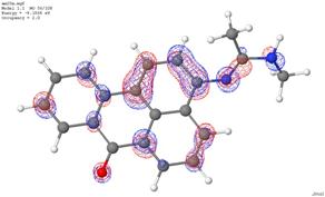
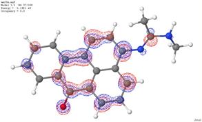
Benzantrona atvasinājumu elektroniskie spektri tika aprēķināti izmantojot ZINDO/S metodi. Aprēķini liecina par intensīvu (oscilators spēks ir ~ 0,6-0,9) absorbcijas joslu pie 370-380 nm. Pamatā tas ir elektronu pāreja HOMO/LUMO.
Benzantrona atvasinājumu fizikāli-ķīmisko īpašību un ķīmiskās struktūras korelatīvā analīze
V. Savenkovs1, I.Gotlibovičs1
1Latvijas Universitātes Mikrobioloģijas un biotehnoloģijas institūts, Rīga, Latvija;
e-pasts: newmikrob@latnet.lv
Dotā projekta ietvaros veidotā fluoroforu datu bāze atrodas pastāvīgā attīstībā. Ar katriem nākamajiem jaunajiem eksperimentālajiem datiem tiek uzstādīti jauni uzdevumi viņu apkopojumam. Lai atrisinātu tādus uzdevumus nepieciešams piesaistīt adekvātas matemātiskās apstrādes metodes, kas pie pietiekoša eksperimentālo datu apjoma var palīdzēt apkopot šo datu masīvu ar sekojošu to izmantošanu saistības meklēšanai starp pētāmo savienojumu dažādiem parametriem.
Atrastās saistības tālāk var kalpot par pamatu jaunsintezējamo fluoroforu to vai citu īpašību modelēšanai. Šajā datu bāzes attīstības etapā ir aprēķināti fluoroforu molekulu ģeometriskie parametri, kā arī veikta korelatīvā analīze starp absorbcijas maksimuma nobīdes lielumiem un emisiju rindai jaunsintezēto benzantrona atvasinājumu, kas parādās atšķirīgos pēc polaritātes pakāpes šķīdinātājos ar šo savienojumu atsevišķu atomu lādiņu lielumiem (AM1 un Zindo/S modeļi).
Augu šūnu funkcionālā stāvokļa pētīšana, izmantojot jauniegūtus fluoroforus
N. Škute
Ecoloģijas Instituts, Daugavpils Universitāte, 13 Vienibas , LV-5401, Daugavpils (Latvia);
e-mail: natalja.skute@inbox.lv
Šūnu bioloģijā ļoti strauji attīstās gan tradicionālās (piemēram, izmantojot ELISE), gan modernās (pielietojot daudzkrāsu proteīnus) fluorescentās analīzes metodes. Jaunu fluorescentu zonžu atklāšana dod būtisko impulsu dažādos šūnu bioloģijas pētījumu virzienos. Īpaša nozīme ir fluorescento zonžu atklāšanai, kuras būtu specifiskas noteiktu šūnu sastāvdaļu iekrāsošanai un kvantitatīvai nukleīnskābju, olbaltumvielu u.c. bioloģiski svarīgu vielu analīzei, kuras nebūtu toksiskas un izmantojamas pētījumos in vivo.
Lai noskaidrotu jaunsintezēto fluoescento vielu toksiskumu tika analizēta šo vielu ietekme uz antioksidantu fermentu izoformu veidošanos, pigmentu koncentrāciju, hlorofila absorbciju un fluorescenci dažādos augu orgānos [1, 2]. Tika analizēta projektā sintezēto vielu ietekme uz vesela auga orgāna augšanu, izmantojot tabakas lapu audu kultūru un kviešu dīgstu modeli, kā arī ietekme uz augu šūnu dzīves ciklu, dažādu augu šūnu organoīdu uzbūve un tās izmaiņas 3D [4]. Tika veikti pētījumi par augu šūnu fizioloģiskā stāvokļa izmaiņām dažādos apstākļos (siltumšoks, sāļu stress) [2, 3]. Tika pētītas morfoloģisko parametru izmaiņas, augu šūnas membrānu caurlaidības izmaiņas funkcionāli dažādos kviešu dīgstu orgānos siltuma šoka ietekmē. Tika veikti antioksidantu enzīmu elektroforetiskās aktivitātes pētījumi un lipīdu peroksidācijas produktu analīze [1]. Lai noskaidrotu mitohondriju lomu oksidatīvajos bojāejas procesos tika veikti eksperimenti par ciklosporīna un antimicīna lomu šajos procesos.
1. J.Orlova, N. Škute. Changes of fluorescence emission spectrum in first leaf and coleoptile cells of wheat seedlings under light stress. 6th International Conference ‘Research and Conservation of Biological Diversity in Baltic Region’, Daugavpils, 2011, 28-29 April.
2. A.Petjukevičs, N. Škute. Current approaches for bioindication, modeling and salt stress analysis in plants. 6th International Conference ‘Research and Conservation of Biological Diversity in Baltic Region’, Daugavpils, 2011, 28-29 April
3. M.Savicka, N.Škute. Effects of high temperature on malondialdehyde content, superoxide production and growth changes in wheat seedlings (Triticum aestivum L.) 2010. Ekologija. 56 (1–2),. 26–33.
4. M. Selga, M. Selga, A. Ozoliņa. Kodola DNS 3D uzbūves un DNS novietojuma izmaiņas šūnas novecošanas laikā. Daugavpils Universitātes 53. starptautiskā zinātniskā konference, Daugavpils 2011. gada 13.-15. aprīlis.